Journal of
Clinical & Medical Surgery
Clinical & Medical Surgery
www.jclinmedsurgery.com
ISSN 2833-5465
Open Access
Volume 5
Open Access
Volume 5
Shamsiyev Jamshid*; Ruziev Jasur; Shamsiev Azamat; Makhmudov Zafar; Shamsiev Rustam
*Corresponding Author: Shamsiyev Jamshid
Specialized Pediatric Surgeon Clinic, Samarkand State Medical University, Samarkand, Uzbekistan.
Email: shamsiyevja@mail.ru
Article Info
Received: Dec 24, 2024
Accepted: Jan 29, 2025
Published: Feb 05, 2025
Archived: www.jclinmedsurgery.com
Copyright: © Jamshid S (2025).
Abstract...
An analysis was made of 1355 children with a suspected Foreign Body of the Respiratory Tract (FBRT), treated in the Specialized Pediatric Surgeon Clinic of Samarkand Medical University over the past 20 years. Among these patients, the diagnosis was confirmed in 948 patients. According to the method of treatment 948 patients were divided into two subgroups. The first subgroup of 478 patients with FB RT, in the period from 2000 to 2009, the removal of FB was performed using rigid bronchoscopy. The second subgroup of 470 patients treated in the period from 2010 to 2019, the removal of FB was performed by video bronchoscopy. The use of video bronchoscopy contributed to a clear visualization of all departments of the FBRT, made it possible to easily and quickly remove the latter, significantly improved the course of the post-bronchoscopy period and reduced the development of late complications.
Keywords: Foreign body of the respiratory tract; Children; Video bronchoscopy.
Citation: Jamshid S, Jasur R, Azamat S, Zafar M, Rustam S. Diagnostic and Treatment of Children with Foreign Bodies of the Respiratory Tracts. J Clin Med Surgery. 2025; 5(1): 1183.
Introduction
One of the common pathologies in pediatric surgery are Foreign Bodies of the Respiratory Tract (FBRT) manifested in the form of acute respiratory disease or respiratory failure requiring emergency medical care. The annual rate of hospitalization of children with FBRT relation to all children receiving inpatient treatment ranges from 2.7% to 14.3% and depends on the profile of the hospital [2,3,7,10].
Foreign Body (FB) aspiration in the vast majority of cases occurs in childhood (66.1-97.2%), as described in several studies [1,4,6,11,13] and majority in children aged 1-5 years old [3,8,10,12] while a considerable group within 1-3 years of age. The risk factor is the time when children actively start to learn and explore about the environment around, to put their hands into the mouths, learn to chew and swallow solid food, all acts able to enhance the personal sensations.
This problem is relevant in children due to the anatomical and physiological features that contribute to the rapid appearance and development of difficult changes in the Respiratory Tract (RT) that appear when FB enters the tissues of the tracheobronchial tree, and as a result of the appearance of impaired conduction of the RT, the development of bronchopulmonary complications [1,3,10,12].
According to the all-authors opinions [2,3,9,14], an important, most informative method for diagnosing and removing FBs from the RT is diagnostic bronchoscopy, which allows visual examination of the RT. Improvements of respiratory optical trachea-bronchoscopes had improved ability of resolution as much as possible and diagnostic value of endoscopy. The endotracheal tube is most commonly used as the airway for fiberoptic bronchoscopy. However, a laryngeal mask can also be applied for this purpose [5,11,15], which is a kind of compromise between a face mask and an endotracheal tube.
Aim of our study is to improve the results of diagnosis and treatment of foreign bodies in the respiratory tract in children.
Material and methods
From January 2000 to June 2019, 1355 children with suspected FBRT were hospitalized and examined in the Department of Thoracic Surgery of the Specialized Pediatric Surgeon Clinic of SamSMU. Among 1355 patients who were hospitalized with a diagnosis of FB RT in 407 (30.0%) this disease was excluded, and in 948 (70.0%) the diagnosis was confirmed.
The total number of patients - 1355, were divided into two groups:
Clinical comparison group (CCG)
407 patients hospitalized in the period from 2000 to 2019 with an initial diagnosis of FBRT that was subsequently excluded. the patients was admitted with a suspicion of a foreign body according to the parents, who were not confirmed after the examination.
Main clinical group (MCG)
948 patients who were hospitalized in the period from 2000 to 2019, in whom the diagnosis of FBRT was confirmed.
The MCG was subdivided in 2 subgroups:
Subgroup I: 478 patients in which the removal of FB was carried out using rigid bronchoscopy.
Subgroup II: 470 patients in which the removal of a FB was performed using a video-bronchoscopy, ozonization method of the tracheobronchial tree, the introduction of acetylcysteine there and strict indications for repeated sanitation.
Table 1: Distribution of patients according to the age and groups.
| Age | Clinical group | Total | |
|---|---|---|---|
| MCG | CCG | ||
| Up to 1year | 92 (22,6%) | 53 (5,6%) | 145 (10,7%) |
| 1-3 year | 190 (46,6%) | 573 (60,4%) | 763 (56,3%) |
| 3-7 year | 80 (19,7%) | 212 (22,4%) | 292 (21,6%) |
| 7-12 year | 35 (8,6%) | 92 (9,7%) | 127 (9,4%) |
| 12-15 year | 8 (2,0%) | 13 (1,4%) | 21 (1,5%) |
| 15-18 year | 2 (0,5%) | 5 (0,5%) | 7 (0,5%) |
| Total | 407 | 948 | 1355 * |
| χ2=88,595; critical value χ2=15,086 (p=0,01); * p<0.01( total) | |||
As showed in Table 1, among all patients, children 1-3 yrs old prevailed with 56.3% (n=763) while the distribution at other age was not significant.
This age is characterized by the fact that the child indepen- dently, on the basis of his own sensations, learns to properly chew and swallow solid food and boys are significantly more at risk then girls (63.3% vs 36.7%) (Table 2).
Table 2: Distribution of patients by gender.

Children from rural areas prevailed over urban ones by 4.5 times (81.7% vs 18.3%) (Table 3).
Table 3: Distribution of patients by place of citizens.
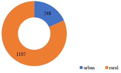
Table 4 presents data on the general condition of patients upon admission.
Table 4: Distribution of patients according to the state of severity at the time of admission.
| Group of patients | Condition of the patient | |||
|---|---|---|---|---|
| Moderate | Severe | Critical | Clinically death | |
| MCGn=407 | 351 (86,2%) | 44 (10,8%) | 12 (3,0%) | - |
| CCGn=948 | 811 (85,5%) | 123 (13,0%) | 12 (1,3%) | 2 (0,2%) |
| total n=1355 | 1162 (85,8%) | 167 (12,3%) | 24 (1,8%) | 2 (0,1%)* |
| χ2=6,508; critical value χ2=7,815 (p<0,01); p=0,090 *p>0,05 | ||||
85.8% of the children were admitted to the hospital in mod- erate clinical condition while only 12.3% were in severe condi- tion and a minor group of 1.8% in critical condition (Table 4). Only 0.1% arrived at our hospital clinically death
Season flu was not a risk for the admission of patients with suspected FBRT, although there is an increase in admission in May, September and October (Table 5) similar for the overall admission of patients with FB Group, while in the MCG, a slight increase was observed from August to December.
Table 5: The frequency of hospitalization of patients with suspected FB RT depending on the season of the year.

In MCG subgroup I, a flexible bronchoscope, proximal illu- mination, Friedel systems (Germany) were used. In the MCG Subgroup II, in order to extract the FB RT, the respiratory video bronchoscope EndoPik (South Korea) was used. In this sub- group, during bronchoscopy, the tracheobronchial tree was washed with ozonized saline, and at the end, an acetylcysteine solution was injected.
Results
In a comparative analysis of the quality of the view of the respiratory tract using a Friedel bronchoscope and a respiratory video bronchoscope, the latter turned out to be more effective. So, when considering the quality of the review of the trachea, main bronchus, lobar bronchi and segmental bronchi, a good (4 points) value of these indicators in subgroup II of the MCG (n = 470, 532 bronchoscopy) was 100% for each section of the TBT, while in subgroup I of the MCG (n=478, 649 bronchoscopies) they were 46.2%, 17.9%, 0% and 0%, respectively.
The average value of FB visibility quality in subgroup II MCG was 3±0.0, while in subgroup I MCG this indicator was 1.92±0.02. In the subgroup II of the MCG, the criterion “FB capture” was on average 3.96±0.01, and in the subgroup I of the MCG - 2.67±0.02.
According to the criterion “injury to the walls of the bronchi”, the average value of which in subgroup I and subgroup II of the MCG was 2.38±0.03 and 3.61±0.03 points, respectively, videobronchoscopy was significantly less traumatic.
Table 6: The frequency of bronchoscopy in one patient in MCG subgroup.
| The frequency of bronchoscopy | IInd Subgroup MCG 470 (455)* | Ist MCG 478(459)* |
|---|---|---|
| Once | 383 (81,7%) | 298 (62,4%) |
| Twice | 68 (14,5%) | 134 (28,0%) |
| Three times | 3 (0,6%) | 25 (5,2%) |
| Four times | 1 (0,2%) | 2 (0,4%) |
| Notcarried out | 15 (3,0%) | 19 (4,0%) |
| Total bronchoscopies | 532 | 649 |
| χ2=50,722; critical value χ2 (p=0,01) =13,277; p<0,001 | ||
Note: * - in subgroup II MCG, out of 470 patients, bronchoscopy was performed in 455 (15 children with self-terminated FB TR showed no indications for bronchoscopy); ** - in subgroup I MCG, out of 478 patients, bronchoscopy was performed in 459 patients (19 children after self-removal of FB RT did not undergo bronchoscopy due to lack of indications).
The duration of bronchoscopy during the removal of FBs from the RT was significantly less when using a video bronchoscope and amounted to 14.45±0.1 minutes versus 21.39±0.4 minutes when using the Friedel bronchoscope. The subgroups were also evaluated in a comparative aspect by the duration of the period of clinical manifestations after bronchoscopy, the absence or presence of complications, the frequency of bronchoscopy, the number of surgical removal of FBs from the RT, the time from the start of treatment to recovery.
The frequency of performed bronchoscopies for FB RT and their complications in children until complete recovery is shown in Table 6.
As shown in Table 6, the number of bronchoscopies in subgroup II was significantly lower than in subgroup I. Differences in treatment effectiveness is significant (χ2=50.722; critical value χ2 (p=0.01) =13.277; p< 0.001). Similarly, in subgroup I, bronchoscopy was performed in 459 out of 478 patients, 19 patients had self-expulsion of the foreign body. 459 patients of this group underwent 649 bronchoscopies: 298 (62.4%) once, 134 (28.0%) twice, 25 (5.2%) three times and 2 (0.4%) four times. 161 (33.6%) patients required repeated sanitation.
In all patients, during videobronchoscopy, the airways were completely sanitized and specific indications were set for repeated medical-sanitation bronchoscopy. And in the subgroup of patients with the Friedel bronchoscope in 12 (2.5%) children, it was not possible to remove the FB completely or it was not found during the first manipulation.
Bronchoscopy was repeated trice in 3 pts (0.6%) of subgroup II, while in subgroup I in 25 (5.2%). 4 procedures were performed in Subgroup I (2 -0.4%) and in 1 (0.2%).
The course of the post-bronchoscopy period based on the clinical manifestations of the disease is shown in Table 7.
Table 7: Duration of clinical manifestations of the course of the postoperative period in subgroups of the main clinical group (in days).
| Indicators | I subgroup of the mainclinical group (n=478) | IIsubgroup of the main clinical group (n=470) |
|---|---|---|
| The duration of the cough | 13,0±0,2 | 10,5±0,1 |
| Recovery period of vesicular (puerile) breathing | 12,0±0,2 | 8,5±0,1 |
| Wheezing duration | 10,0±0,2 | 7,0±0,1 |
| The differences arestatistically significant (p=0.000000); Number of de- grees of freedom f = 946; Critical value of Student's t-test =1.972, at signifi- cance level α =0.05 | ||
Table 7 shows that in subgroup II of the MCG, the duration of the cough period averaged 10.5±0.1 days, while in subgroup I it reached 13.0±0.2 days. In subgroup II MCG during auscultation, the recovery time of vesicular (puerile) breathing was at the level of 8.5±0.1 days, and in subgroup I - 12.0±0.2 days. Wheezing during auscultation in I and II subgroups of MCG persisted for 10.0±0.2 and 7.0±0.1 days, respectively.
So, in the subgroup using a video bronchoscope, there was a decrease in the duration of clinical manifestations in the post-bronchoscopy period, in particular, the duration of the cough period by 2.5 days, the recovery period of vesicular or puerile breathing by 3.5 days and the duration of wheezing by 3 days. The differences obtained in the calculations were significant.
In our sample, we obtained a 95.1% positive history of aspiration confirmed by bronchoscopic examination.
Out of 1355 patients admitted to our clinic with suspected FB RT, bronchoscopy was performed in 1153 (85.1%) patients, including 914 (96.4%) children of the main clinical group (459 (96.0%) in subgroup I and 455 (96.8%) in subgroup II) and 239 (58.7%) children of the clinical comparison group. In 34 children of the MCG group, bronchoscopy was not performed, since there were clear anamnestic data on self-discharge of FB and there were no indications for manipulation. In all 239 cases of bronchoscopy in CCG, no foreign body or signs of its presence in the respiratory tract were found. In all 914 MCG patients who underwent bronchoscopy, FBs were removed or had signs of being in the tracheobronchial tree.
Discussion
The data presented, clearly demonstrate the advantages of optical bronchoscopy for the fine visualization of all parts of the tracheobronchial tree and identify not only the foreign object with its size, shape, edges, etc.
It is also possible to clearly determine the nature, depth and prevalence of injury of the trachea and bronchi as a result of exposure to FB allowing excellent conditions to visualize, capture and extract it. In addition is possible to sanitize the trachea-bronchial tree, and also to repeated medical and sanitation procedures. In subgroup I of the MCG (2000-2009), not having such opportunities, it was not possible in all cases to fully sanitize the bronchi, and the indications for repeated bronchoscopy were based only on clinical data.
In order to control the course of the post-bronchoscopy period and the effectiveness of the treatment, daily monitoring of clinical symptoms was carried out. The patient’s body temperature and its duration, absence or severity of cough, if it is present (3 points - no cough, 2 points - periodic cough, 1 point - single cough); the absence or severity of wheezing, if it presents during auscultation of the lungs (3 points - no wheezing, 2 points - single wheezing, 1 point - widespread multiple) and the recovery period of vesicular (puerile) breathing in days.
Conclusion
In conclusions, based on the data of our study, video bronchoscopy contributes to a clear visualization of all parts of the respiratory tract and the aspirated object, allows you to easily and quickly remove the latter from the TBT, significantly improves the course of the post-bronchoscopy period and prevents the development of late complications.
References
- Akopov AL, Molodtsova VP, Chistyakov IV, Ilyin AA, Vasil’eva MA. A rare case of an undiagnosed foreign body in the bronchus. Bulletin of Surgery. 2015; 174: 82-85.
- Dyakonov VL. Issues of urgent therapy for foreign bodies of the bronchi in children: Abstract of the diss-candidate of medical sciences. Samara. 1993: 24.
- Kajina VA, et al. Removal of foreign bodies from the tracheobronchial tree in children of the Grodno region: 10-year experience of rigid bronchoscopy with video imaging. Journal of the Grodno State Medical University. 2015: 108-113.
- Kugaevskiy VN, Bocharnikov ES, Poleshchuk VV, Ponomarev VI. Ten years of experience in helping patients with foreign bodies in the respiratory tract. Basic research. 2012: 284-288.
- Mustafaev DM, Egorov VI. Unusual foreign bodies of the respiratory tract in children // Medicine of Extreme Situations. 2018; 20: 115–120.
- Rusetsky Yu Yu, Spiranskaya OA, Chernishenko IO. Foreign bodies of the lower respiratory tract in children: modern diagnostic and therapeutic approaches. Pediatrics. 2015; 94: 30-35.
- Spiranskaya OA, Rusetsky Yu Yu, Chernyshenko IO. The use of optical respiratory bronchoscopy in the removal of foreign bodies in the lower respiratory tract in children. Otorhinolaryngology - Bas, Moiyn Khirurgiasy. 2011: 93-94.
- Kharitonova A Yu, Shavrov AA, Kalashnikova NA, Shavrov AA. (ml.). Diagnostic bronchoscopy in children. Questions of modern pediatrics. 2013; 4: 112-119.
- Shamsiev JA, Ruziev JA, Zainiev SS, Isakov AM. Foreign bodies of the respiratory tract in children // Science and practice: implementation to modern society. 2020: 378–383.
- Boufersaoui A, Smati L, Benhalla KN, Boukari R, Smail S, Anik K, et al. Foreign body aspiration inspiration children: experience from 2624 patients. Int J Pediatr Otorhinolaryngol. 2013; 77: 1683-8.
- Ding G, Wu B, Vinturache A, Cai C, Lu M, Gu H. Tracheobronchial foreign body aspiration in children: A retrospective single-center cross-sectional study. Medicine (Baltimore). 2020; 99: e20480.
- Kapoor R, Chandra T, Mendpara H, Gupta R, Garg S. Flexible Bronchoscopic Removal of Foreign Bodies from Airway of Children: Single Center Experience Over 12 Years. Indian Pediatrician. 2019; 56:560-562.
- Kogure Y, Oki M, Saka H. Endobronchial foreign body removed by rigid bronchoscopy after 39 years. Interact Cardiovasc Thorac Surg. 2010; 11: 866-68.
- Korlacki W. Foreign body aspiration and therapeutic role of bronchoscopy. Pediatr Surg Int. 2011; 27: 833–837.
- Aydogan LB, et al. Rigid bronchoscopy for the suspicion of foreign body in the airway. Int J Pediatr Otorhinolaryngol. 2006; 70: 823.