Introduction
Implant placement has become a common procedure when
one has to rehabilitate missing teeth functionally and aesthetically. With better understanding and advances in technology
there are many procedures available to rehabilitate maxillary
arch and clinicians have many options of bypassing the sinus
such as zygoma implants, all-on-4 technique, indirect snus lift
or graft less solutions and short implants. However, each aforementioned technique is not as predictable as direct sinus lift
is specially in cases of severely atrophic maxilla [1,2]. Short
implants have less bone-to-implant contact and have more
chances of failure in load bearing areas. In a meta-analysis by
Papaspyridakos et al [3], authors stated that short implants (≤6
mm) have less predictable survival rates compared to longer
implants (>6 mm) after periods of 1-5 years in function. When
it comes to indirect sinus lift, the procedure does not increases
the height of maxillary sinus sufficiently [4], leaving direct sinus
lift a predictable treatment choice. However, many patients are
anxious in maxillary sinus grafting and the complications which
follow the surgery, hence procedures like all-on-4 were developed which can rehabilitate the entire arch along with bypassing the sinus.
In order to do so, knowing anatomy and the arterial blood
supply becomes paramount. Structure wise, Maxillary Sinus
(MS) is pyramidal in shape and largest of the paranasal sinuses
[5]. The anterior wall of the MS is formed by the facial surface
of the maxilla. and is internally grooved by the canalis sinuosus
(which houses the anterior superior alveolar nerve and vessels) [5]. It receives blood supply via maxillary artery and its
branches. In this particular paper, the artery of interest is Alveolar Antral Artery (AAA) which is an anastomosis of the Posterior Superior Alveolar Artery (PSAA) and the Infraorbital Artery
(IOA) [6]. As the name suggests it appears on the maxillary facial
plate near to or in close approximation with maxillary second
premolar and first molar area, a position from where it takes a
U-turn, roughly paralleling the sinus floor. Not much has been
reported regarding this artery in the literature as it goes undetected and the clinician only comes to know when it interferes
during osteotomy (10-30% of cases) or any complication [7,8].
It has higher detection rates in males due to larger diameters,
as well as in narrower maxillary sinuses <14 mm in width. Immediate complication is bleeding, which can be called as the
‘gateway complication’ further leading to a series of complications impact treatment prognosis and outcome and or abortion of the surgical procedure [6,9]. Also the vertical distance of the
artery from the alveolar ridge varies in patients depending upon
the resorption of the ridge. Solar et al [10] reported a range of
15 - 25 mm of vertical positioning of the artery in alveolar bone
in a mixed dentate/edentulous population. However a more reliable measurement would be the one done via CBCT, where
ridge resorption will not influence the vertical distance had
been found to be mean of 7.66 mm [11]. Park et al [12] found
this height to be between 7.71 - 8.01 mm, and suggested a lateral window of 8mm for visualization, instrumentation and graft
placement if sinus lift is to be performed. Studies have shown
that the artery has an average diameter of 1.5 mm [13], and
that a Diameters < 0.5 mm does not any significant bleeding to
interfere with surgery [14]. But Testori et al [15] suggested that
a small calibre vessel on CBCT, may correspond to a much larger
calibre vessel clinically.
Case report
A 59-year-old male patient reported to the dental office
with missing maxillary teeth due to poor oral hygiene. Treatment planning was done for all on four implant placement followed by prosthesis. A complete medical history was obtained
and was negative for any significant medical problems. Patient
denied being allergic to any medication as well. Patient agreed
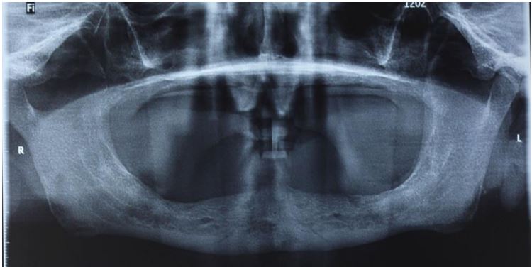
for the implant placement and was advised orthopantomogram
(Figure 1). On the day of the surgery, before commencing the
procedure under strict asepsis, patient was asked to rinse with
0.12% chlorhexidine gluconate mouthwash (Peridex; Proctor &
Gamble, Cincinnati, OH). Local anaesthesia with a vasoconstrictor was infiltrated buccally and palatally into the posterior and
anterior maxilla on both the sides and Using a S-blades (straight)
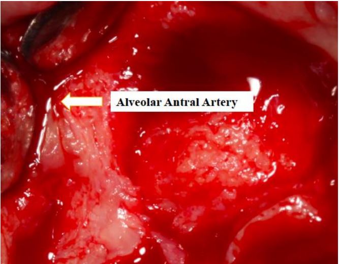
(Zabby, India) incision was given on the crest of the ridge in the
region of 15-25. While giving the incision, bleeding was noticed
in the region of 15 which intensified during the flap reflection
(Figure 2). Bleeding was pulsatile indicating arterial bleed. Initially to control pressure pack and ice pack was given, in the
meantime the bleeder was isolated and the vessel was ligated
(Figure 3). The bleeding could be controlled and the procedure
was completed by placing 4 Bioner implants (Bioner, Spain) of
size 4/10mm. Sutures were placed and patient was kept on basic medication for pain and infection control. Immediately after
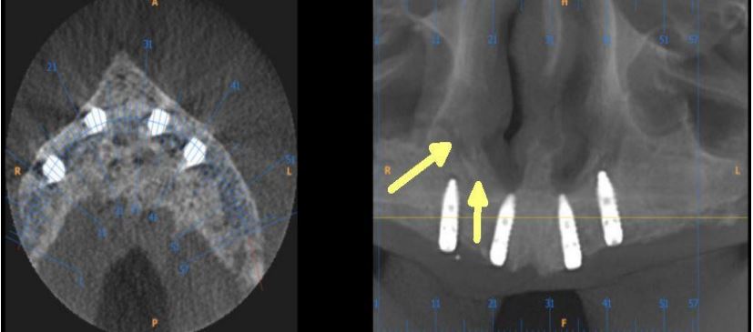
the surgery patient was advised for CBCT, as shown in the figure
4 a coronal view and 4b (yellow arrows) position of the artery
can be seen.
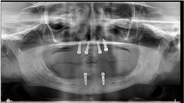
In the follow-up sessions, patients was comfortable and did
complain of mild swelling which subsided with 4-5 days. A postop CBCT showed excellent recovery, bone width and proper implant placement (Figure 5).
Discussion
Encountering AAA complication during all-on-4 procedure
in the region of 14,15 has not been reported so far, however
literature reports bleeding complication from AAA during sinus
lift procedures. Another complication that occurs is infection
in about 3% of the cases in 1% of the cases loss of graft [16],
and this usually happens after heametoma formation. In simple
terms, larger the vessels more the bleeding will be. According
to Ella et al [17] the risk involving AAA in osteotomies can be
>10% whereas according to authors Chan & Wang [18] and
Elian et al [8] it is 20%. Authors Jensen et al [19] have reported severe bleeding in sinus elevation surgery through transcrestal
approach, where in, bleeding let to swelling and consecutive
hospitalization for 3 days after which normal functioning was
regained. Hence management of the complication plays an important part for the clinician.
The first and foremost is the application of pressure pack and
ice pack to control the bleeding along with topical thrombin.
Other products such as SURGICEL® Absorbable Hemostat, SURGICEL™ Powder (Surgicel; Johnson & Johnson Co., Somerville,
NJ), Bone-wax can also be used. Usually because of bleeding at
the surgical sight, pinpointing the exact bleeding spot becomes
difficult but if that can be isolated then Electrocautery/ chemocautery or ligation can be done. As aforementioned, if the vessel is large and bleeding cannot be controlled, the procedure
should be aborted and patient should be hospitalised. Use of
piezoelectric devices safely bypasses the vessels as it only cuts
the bony surface, avoiding any chance of vessel rupture. The
only disadvantage is that it is time-consuming method, but better when it comes to any complication or aborting the procedure. In a surgical double-window technique described by Maridati et al [20] osteotomy is made above and below the vessel
leaving a thin bridge of bone holding the vessel intact. However this is a difficult technique to follow and does not work
in terms of instrumentation, implant placement and septated
sinuses [6]. The simplest of the methods to avoid this complication is detection which is best achieved with CBCTn
, however
even with CBCT, AAA can be detected only in 50% of the cases
[21,22], (may be because the vessel is too small to be detected
by CBCT) which does not mean that there is an absent vessel or
anastomosis does not exist, cadaveric studies have shown that
anastomosis is present 100% of the time [23]. But it goes undetected or unreported, simply because many clinicians assume
that the anastomosis does not exist or use basic radiographic
techniques for the implant placement.
In the case discussed here, authors encountered a small vessel wherein the bleeding was easily controlled with pressure
packs and ice packs. The site of implant placement is a safe zone
and such bleeding complication usually occur during sinus lift
procedures and not in the anterior region. A simple detection
could have helped clinicians to plan osteotomy better, fortunately the vessel was not big the bleeding could be controlled
by ligation, otherwise, may be the implant placement had to be
postponed. This proves, CBCT is an excellent tool and should be
used more often for the case planning and detection of pathologies [21].
Conclusion
Dental radiographs are an important tool in accurate diagnosis and treatment planning [21]. It is also the most common
and important investigation carried out before any dental procedure which requires surgical or corrective intervention [21].
Thus, accuracy of the x-ray taken whether it is an Intraoral Periapical Radiograph (IOPA) or an Orthopantomogram (OPG) becomes paramount. However, these investigative tools provide a
2D image of a 3D object and are subject to false positive errors.
Also, patients should be counselled and motivated for proper
investigations. Above all, the surgeon must be competent to
anticipate and effectively manage complications encountered
during the surgery, as we could in the case discussed.
Acknowledgement: None.
Conflict of interest: None to declare.
References
- Chipaila N, Marini R, Sfasciotti GL, Cielo A, Bonanome L, et al. Graft less Sinus Augmentation Technique with Contextual Placement of Implants: A Case Report. Journal of Medical Case Reports. 2014; 8: 437. doi.org/10.1186/175219478437.
- Bedrossian E, Rangert B, Stumpel L, Indresano T. Immediate Function with the Zygomatic Implant: A Graftless Solution for the Patient with Mild to Advanced Atrophy of the Maxilla. Int J Oral Maxillofac Impla. 2006; 21: 937-942.
- Papaspyridakos P, De Souza A, Vazouras K, Gholami H, Pagni S, et al. Survival rates of short dental implants (≤6 mm) compared with implants longer than 6 mm in posterior jaw areas: A meta-analysis. Clin Oral Implants Res. 2018; 29(16): 8-20. doi: 10.1111/clr.13289.
- Balaji SM. Direct v/s Indirect sinus lift in maxillary dental implants. Ann Maxillofac Surg. 2013; 3(2): 148-53. doi: 10.4103/2231-0746.119228.
- Standring S. Gray’s anatomy: The anatomical basis of clinical practice. 41st ed. London: Elsevier Health Sciences. 2015.
- Yang D, Lee N. A Simple Method of Managing the Alveolar Antral Artery during Sinus Lift Surgery. Int J Otolaryngology Head Neck Surg. 2021; 10: 131-146. doi: 10.4236/ijohns.2021.103014.
- Lee CYS. Brisk, Prolonged Pulsatile Hemorrhage during the Sinus Graft Procedure: A Case Report with Discussion on Intra-Operative Hemostatic Management. Implant Dent. 2010; 19: 189-195.
- Elian N, Wallace S, Cho SC, Jalbout ZN, Froum S. Distribution of the Maxillary Artery as It Relates to Sinus Floor Augmentation. Int J Oral & Maxillofac Implants. 2005; 20: 784-787.
- Varela-Centelles P, Loira M, González-Mosquera A, et al. Study of Factors Influencing Preoperative Detection of Alveolar Antral Artery by CBCT in Sinus Floor Elevation. Scientific Reports. 2020; 10: 10820. Doi.org/10.1238/s41598-020-67644-9.
- Solar P, Geyerhofer U, Traxler H, Windisch A, Ulm C, et al. Blood Supply to the Maxillary Sinus Relevant to Sinus Floor Elevation Procedures. Clin Oral Implants Res. 1999; 10: 34-44. Doi.org/10.1034/j.1600-0501.1999.100105.x.
- Varela-Centelles P, Loira-Gago M, Gonzalez-Mosquera A, Seoane-Romero JM, Garcia-Martin JM, et al. Distance of the Alveolar Antral Artery from the Alveolar Crest. Related Factors and Surgical Considerations in Sinus Floor Elevation. Medicina Oral, Patologia Oral, Cirugia Bucal. 2016; 21: e758-e765. doi.org/10.4317/medoral.21475.
- Park WH, Choi SY, Kim CS. Study on the Position of the Posterior Superior Alveolar Artery in Relation to the Performance of the Maxillary Sinus Bone Graft Procedure in a Korean Population. J Korean Ass Oral Maxillofac Surg. 2012; 38: 71. Doi.org/10.5125/jkaoms.2012.38.2.71.
- Kim JH, Ryu JS, Kim KD, Hwang SH, Moon HS. A Radiographic Study of the Posterior Superior Alveolar Artery. Implant Dent. 2011; 20: 306-310. doi.org/10.1097/ID.0b013e31822634bd.
- Rysz M, Ciszek B, Rogowska M, Krajewski R. Arteries of the Anterior Wall of the Maxilla in Sinus Lift Surgery. International J Oral and Maxillofac Surg. 2014; 43: 1127-1130. doi.org/10.1016/j.ijom.2014.02.018.
- Testori T, Rosano G, Taschieri S, Del Fabbro M. Ligation of an Unusually Large Vessel during Maxillary Sinus Floor Augmentation. A Case Report. Euro J Oral Implantol. 2010; 3: 255-258.
- Chiapasco M, Casentini P, Zaniboni M. Bone Augmentation Procedures in Implant Dentistry. Inte J Oral Maxillofac Impla. 2009; 24: 237-259.
- Ella B, Sédarat C, Noble RDC, et al. Vascular Connections of the Lateral Wall of the Sinus: Surgical Effect in Sinus Augmentation. Int J Oral Maxillofac Impla. 2008; 23: 1047-1052.
- Chan HL, Wang HL. Sinus Pathology and Anatomy in Relation to Complications in Lateral Window Sinus Augmentation. Impla Dent. 2011; 20: 406-412. doi.org/10.1097/ID.0b013e3182341f79.
- Jensen SS, Eriksen J. Schiodt M. Severe Bleeding after Sinus Floor Elevation Using the Transcrestal Technique: A Case Report. Euro J Oral Implantol. 2012; 5: 287-291.
- Maridati, P, Stoffella E, Speroni S, Cicciu M, Maiorana C. Alveolar Antral Artery Isolation during Sinus Lift Procedure with the Double Window Technique. Open Dentist J. 2014; 8: 95-103. Doi.org/10.2174/1874210601408010095.
- Shukla S, Chug A, Afrashtehfar KI. Role of Cone beam computed tomography in diagnosis and treatment planning in dentistry: An update. J Int Soc Prev Community Dent. 2017; 7(Suppl 3): S125-S136.
- Ilgüy D, Ilgüy M, Dolekoglu S, Fisekcioglu E. Evaluation of the Posterior Superior Alveolar Artery and the Maxillary Sinus with CBCT. Brazilian Oral Res. 2013; 27: 431-437. https: //doi.org/10.1590/S1806-83242013000500007.
- Bernardi S, Mummolo S, Ciavarelli LM, Li Vigni M, Continenza MA, et al. Cone Beam Computed Tomography Investigation of the Antral Artery Anastomosis in a Population of Central Italy. Folia Morphologica (Warsz). 2016; 75: 149-153. doi.org/10.5603/FM.a2015.0095.