Introduction
Foot drop is a condition that makes walking difficult and
interferes with active living [1,2]. Tendon transfers, such as the
tibialis posterior tendon, are the most important adjunctive
procedures in the treatment of foot drop [3,4]. It is important
to apply the increasingly popular tendon transfer techniques
and to understand the functional consequences of the type
of surgical technique [5-7]. In particular, tendon transfer has
been shown to improve patient satisfaction by increasing
mobility and walking independence. In the treatment of foot
drop, tendon transfer over the interosseous membrane and
tendon fixation to bone have been the preferred techniques in
recent years [8-10]. In our case, we used combined tendon-to-tendon and tendon-to-bone fixation, preferring the circumferential route and the superficial retinacular passage. The wide-spread use of these methods is important for the patient’s
participation in active life and satisfaction.
Case presentation
A 53-year-old male patient; with right foot drop following
lumbar spinal surgery 7 years ago, was planned for tendon
transfer. Passive dorsiflexion was 30-35 degrees, so Achilles
lengthening procedure was not necessary. Neurological examination showed dorsiflexion 1/5, thumb extension 1/5, and foot
inversion 2/5. No significant muscle atrophy was observed. In
the surgical procedure, after finding the insertion point of the
posterior tibial tendon through a small incision at the navicular
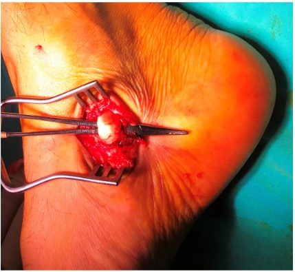
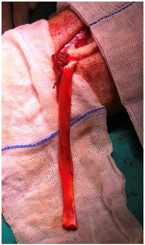
bone, the tendon was released through a second vertical incision 7 cm superior to the medial malleoli (Figures 1 and 2). A
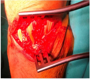
third incision was then made parallel to the 5 cm pili on the dorsal side of the foot, which will dominate the extensor tendons
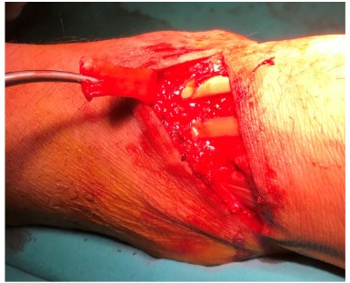
(Figure 3). Using Deschamp, the tendon was transferred dorsally over the extensor retinaculum via the circum-tibial route
(Figure 4). The tendon was then bisected 3 cm distal to the tendon. The medial portion was ligated to the navicular bone and
the lateral portion was ligated to the extensor tendons with the
foot in 20 degrees of dorsiflexion. The patient was started on a
physiotherapy programme after 6 weeks of splinting and at the
6 month follow-up, foot dorsiflexion was recorded as 3/5 and
ROM angle as 45 degrees.
Discussion
Foot drop may be of neurological origin related to central,
spinal, and peripheral nerves or muscular origin such as muscle
injury and compartment syndrome. It causes increased knee
and thigh flexion on the affected side and hip elevation on the
same side during the swing phase of gait, which impairs quality of life. Although gait can be improved to some extent with
a foot-ankle orthosis that prevents further plantar flexion in
the neutral position, surgical treatment should be performed
early before spasticity and muscle atrophy develop. The most
common cause of foot drop is injury to the peroneal nerve. In
this case, early nerve repair is the first option. If this is not sufficient, a tendon transfer should be performed. Posterior tibial
tendon transfer has been performed with remarkable success
since 1933. Although tendon transfer is the gold standard, arthrodesis and tenodesis can also be performed [1,2]. Although
postoperative dorsiflexion strength averages 33%, it improves
function in activities of daily living and ambulation [1,2]. If the
clubfoot is not treated, an equinovarus deformity will develop
over time [3]. During surgery, a small incision is made on the
navicular bone to locate the attachment of the posterior tibial tendon to the tuberosity. A second incision, 7.1 cm above
the medial malleolus in males and 6.4 cm in females, allows
us to obtain the longest mobile tendon without damaging its
origin [4]. The tendon is transferred to the dorsum of the foot
through the 3rd incision parallel to the ankle line. Intraosseous
(transmembranous) and circumtibial routes may be preferred
for transfer. Some argue that the transmembranous technique
results in greater dorsiflexion and ROM angle [5]. However,
the risk of vascular damage and adhesion formation is higher
with this method [6]. As the circumferential tract is longer, the torque for dorsiflexion will be greater. Data from cadaveric
studies have shown that tendon length is insufficient for transmembrane transfer [7]. Another important consideration is the
retinacular junction. Crossing the external retinaculum results
in more effective motion [8].
Fixation can be tendon-tendon or tendon-bone. The recipient tendons are the anterior tibial or anterior tibial and extensor
tendons [9]. Tendon-tendon fixation should be performed with
the foot in 20 degrees of dorsiflexion, allowing for stretching
over time [10]. Tarsal or metatarsal bones may be preferred for
tendon-bone fixation. This is done by opening a bone tunnel.
This causes neuropathic arthropathy. Bone fixation alone will
not prevent the toes and fingers from falling off. After splinting
for 4-6 weeks postoperatively, the physiotherapy programme
is started early. In our case, we used combined tendon-tendon
and tendon-bone fixation, preferring the circum-tibial route and
the superficial retinacular passage. We suggest that combined
fixation should be emphasised in terms of functional outcomes.
Conflict of interest: No potential conflict of interest relevant to this article was reported.
References
- Roukis TS, Landsman AS, Patel KE, Sloan M, Petricca D. A simple
technique for correcting footdrop: suspension tenodesis of
tibialis anterior tendon to distal tibia. J Am Poditar Med Assoc.
2005; 95: 154-6.
- Cho BK, Park KJ, Choi SM, Im SM, SooHoo NF. Functional Outcomes Following Anterior Transfer of the Tibialis Posterior Tendon for Foot Drop Secondary to Peroneal Nerve Palsy. Foot ankle
Int. 2017; 38(6): 627-633.
- Wiesseman GJ. Tendon transfers for peripheral nerve injuries of
the lower extremity. Orthop Clin North Am. 1981; 12: 459-67.
- Thamphongsri K, Harnroongroj T, Jarusriwanna A, Chuckpaiwong B. How to harvest the greatest length of tibialis posterior
tendon for tendon transfer: A cadaveric study. Clin Anat. 2017;
30(8): 1083-1086.
- Wagner E, Wagner P, Zanolli D, Radkievvich R, Radenz G, Guzman
R. Biomechanical Evaluation of Circumtibial and Transmembranous Routes for Posterior Tibial Tendon Transfer for Drop foot.
Foot Ankle İnt. 2018; 39(7): 843-849.
- Andersen JG. Foot drop in leprosy and its surgical correction.
Acta Orthop Scand. 1963; 33: 151-71.
- Pappas AJ, Haffner KE, Mendicino SS. Cadaveric limb analysis
of tendon length discrepancy of posterior tibial tendon transfer through the interosseus membrane. J Foot Ankle Surg. 2013;
52(4): 470-4.
- D’Astous JL, MacWiliams BA, Kim SJ, Bachus KN.Superficial versus deep transfer of the posterior tibialis tendon. J Pediatr Orthop. 2005; 25(2): 245-8.
- Goh JC, Lee PY, Lee EH, Bose K. Biomechanical study on tibialis
posterior tendon transfers. Clin Orthop Relat Res. 1995; (319):
297-302.
- D Soares. Tibialis posterior transfer in the correction of footdrop
due to leprosy. Lepr Rev. 1995; 66: 229-34.


