Introduction
Sebaceous Glandhy Perplasia (SGH) is a common benign
proliferative disease, with the incidence of about 1% [1]. The
pathogenesis of SGH is not fully understood. Old age, male,
immunosuppression, as well as exposure to UV radiation may
be risk factors for the SGH. SGH typically appears as isolated or
multiple, yellow, soft, umbilicated papules. Rare variants of SGH
include cases in linear, zosteriform, diffuse, giant and nevoid
form, even with familial history, and so on which manifestations
are often lead to misdiagnosis clinically. In this case,we present
a special linear sebaceous gland hyperplasia behind the right
ear clinically simulating verrucous nevus. Dermoscopy and RCM
played an important role in the diagnosis of SGH.
Case report
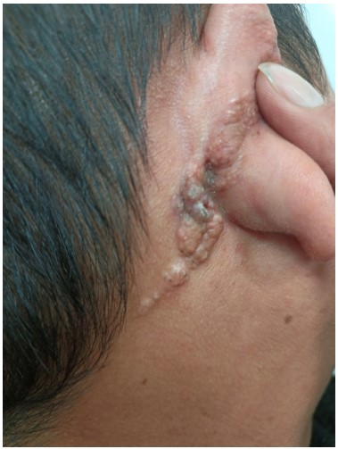
A 40-year-old man suffered from some asymptomatic flesh-colored papules arranged in line behind the right ear for more
than 5 years. The lesions gradually increased and enlarged, with rough and uneven surface. History of prior local trauma or systemic medication was absent. The family history was also destitute. On examination, the flesh-colored to light brown papules
and nodules with a few scales on the surface, localized behind
the right ear involved the auricle in roughly linear distribution,
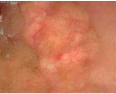
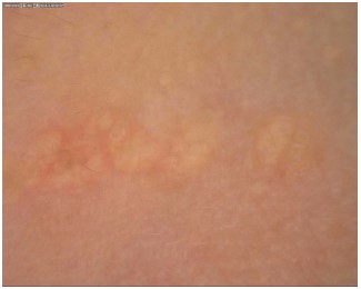
about 2.5 cm × 8 cm in size (Figure 1A). The primary clinical diagnosis was verrucous nevus. Dermoscopy showed a yellowish
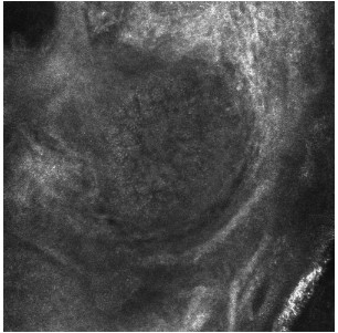
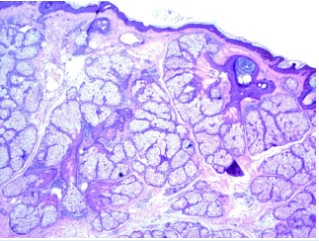
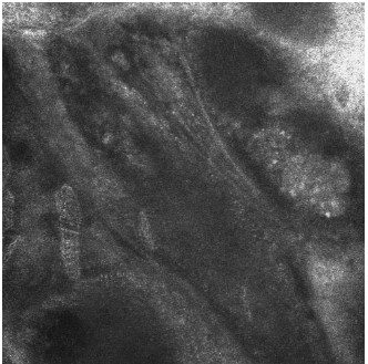
lobulated structure surrounded by crown-shaped vessels (Figure 1B). RCM revealed morula-shaped sebaceous gland structures are seen in the dermis (Figure 1C). Further the biopsy was
taken from the lesions and the histopathology showed that a
large number of mature sebaceous lobules distributed in clusters, with partial catheter malformation but well-differentiated
cells in the dermis, and lymphocytes and histiocytes in small
pieces infiltrated around the capillaries in the superficial dermis
(Figure 1D). The diagnose of line sebaceous gland hyperplasia
was made. The patient refused to treat by surgical or laser excision. After 6 months of follow-up, there was no progress in skin
lesions.
Discussion
SGH is a common benign proliferative skin disorder, mainly
affected adults of middle age. It is caused by the enlargement
of the normal sebaceous glands, but its pathogenesis is unclear.
Usually it presents as multiple, soft, small, yellowish papules
with central umblication, which easily affects forehead, nose
and cheeks [2]. Rare variants of SGH include a linear form, zosteriform arrangement, a diffuse form, a giant form, a nevoid
form, and a familial form Linear form cases reported in the past
presented as small in size, exhibited a linear arrangement on
the supra- or subclavicular areas [3]. Sato et al [4] reported two
cases with linear SGH on the chest displayed as sporadic papules but distributed along the Blaschko line. Another case with
a linear hyperkeratosis plaque mimicking a wart over the right
ear was reported [5]. Our case characted by rough warty plaque
behind the right ear mimicking verrucous nevus clinically, which
is a special clinical manifestation. Diagnosis of SGH depends on
histopathological examination, which characterized by enlarged
sebaceous glands with lobules containing mature sebaceous
cells opening into widened ducts. Dermoscopy and RCM are
non-invasive techniques developed in recent years to observe
the microscopic morphological characteristics of epidermis and
superficial dermis. The typical dermoscopy character of SGH
is well-demarcated yellow-white lobular structure surrounded
by crown-shaped vessels, which is a characteristic feature to
make a diagnose of SGH. RCM shows a lobulated proliferation
composed of round cells with bright speckled cytoplasm and
a centrally located nucleus as “morula-shaped”, and a dilated
central follicular infundibulum at the level of papillary dermis[6].
Dermoscopy and RCM are helpful for us to make a diagnosis of
SGH.
Typical sebaceous hyperplasia is easy to be diagnosed, while
SGH with special manifestations should be distinguished from
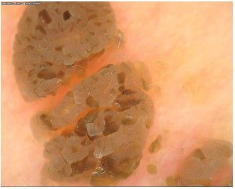
sebaceous nevus [7], linear epidermal nevus [8] and colloid milium. Under dermoscopy, the sebaceous nevus present with yellow round and oval structures of different sizes, which are unrelated to hair follicles, and accompanied by telangiectasia (Figure
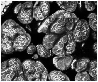
2A). With RCM, the superficial dermis showed grape cluster sebaceous structures, with tubular or stipe structures in the center, and sebaceous lobules with frog egg hyperplasia clustered
in the periphery. The upper epidermis often presented papillomatous hyperplasia (Figure 2B). The linear epidermal nevus
under dermoscopy showed skin color, brown-yellow, brown-brown or brown-black cerebellar gyrus structure and corn keratoid structure (Figure 3A). RCM showed excessive epidermal
keratosis, irregular extension of epidermal processes, papillomatoid hyperplasia, and increased pigment in the basal layer
(Figure 3B). Under the dermoscopy colloid milium show brown-yellow unstructured masses were separated by white stripes
and presented cross-sectional changes of orange petals. There
are branched vessels in and around the septa of the mass [9].
SGH mainly affects the appearance and has a low rate of
cachexia. Therefore, the choice of treatment methods should
take into consideration the patient's wishes and side effects.
Treatments about SGH include topical chemical treatments,
cryotherapy, photodynamic therapy, electrotomy, laser therapy,
oral isotretinoin and surgery [10]. Pigmentation, scarring and
relapse are common adverse reactions.
Funding sources
Chongqing Science and Health Joint Medical Research
Project:2021MSXM118.
References
- Farci F, Rapini RP. Sebaceous Hyperplasia. In: StatPearls [Internet]. Treasure Island (FL): StatPearls Publishing. 2022.
- Gorski K, Korczynska L, Spiewankiewicz B, et al. Vulvar sebaceous
hyperplasia - a problematic dermatosis of the vulva. Ginekol Pol.
2023.
- Franco G, Donati P, Muscardin L, Maini A, Morrone A. Juxta-clavicular beaded lines. Australas J Dermatol. 2006; 47: 204-5.
- Sato T, Tanaka M. Linear sebaceous hyperplasia on the chest.
Dermatol Pract Concept. 2014; 4: 93-5.
- Nair PA, Diwan NG. Sebaceous Hyperplasia Mimicking Linear
Wart over Ear. Int J Trichology. 2015; 7: 170-2.
- Shahriari N, Grant-Kels JM, Rabinovitz H, et al. Reflectance confocal microscopy: Diagnostic criteria of common benign and malignant neoplasms, dermoscopic and histopathologic correlates
of key confocal criteria, and diagnostic algorithms. J Am Acad
Dermatol. 2021; 84: 17-31.
- Jiang Qian, Chen Hongying, Ma Ling, et al. Characteristics of
sebaceous nevus using dermoscopy and reflection confocal microscopy. Chinese Journal Dermatology. 2018; 51: 523-525.
- Meng Ru-song, Cui Yong. Multimodal Dermatologic image Diagnostic Spectrum [M]. People’s Medical Publishing House. 2021;
436-437.
- Ji Ying, Ma Yan, Zhang Jinyu, et al. Dermoscopic observation of
adult colloid milium. Journal Clinical Dermatology. 2020; 049:
29-32.
- Lama, Hussein, Conal M, Perrett. Treatment of sebaceous gland
hyperplasia: A review of the literature.The Journal of dermatological treatment. 2021; 32: 866-877.
