Introduction
Mature cystic teratomas of ovary are most common benign
ovarian neoplasm of reproductive age group [1]. It accounts
for 10-20% of all ovarian neoplasms [2]. Most of them are asymptomatic or may present with complaints of abdominal pain
when they enlarge. Mature cystic teratomas most commonly
present as unilateral cyst, bilateral presentation of mature cystic teratoma is rare and accounts for only 10-15% [3]. Malignant
transformation of mature cystic teratomas seen in 0.1-0.2% of
cases.
Case presentation
We present a case of 35-year multiparous women who
presented with complaints of abdominal pain for 6-7 months.
Ultrasound revealed bilateral cystic lesions of ovary. MRI pelvis showed well circumscribed, lobulated, heterogenous, predominant fat signal intensity lesions in bilateral adnexa. Left
adnexal lesion is larger and measures 13.8 x 9 x 7 cm, smaller
right adnexal lesion measures 9.3 x 7 x 7 cm. Few thick internal
septae with focal internal soft tissue nodules identified in both
adnexal lesions. Imaging features are highly suggestive of bilateral adnexal dermoid cysts. Patient underwent total abdominal
hysterectomy with bilateral salpingo-oopherectomy and specimen was sent for histopathological examination. Intraoperative
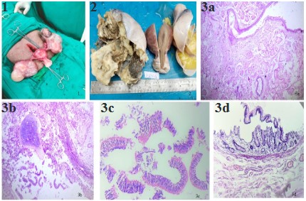
picture of bilateral cystic ovarian masses in shown in Figure 1.
We received hysterectomy specimen with attached bilateral fallopian tubes and bilateral cystic ovarian masses. On gross examination uterus with cervix measured 8.5 x 5 x 4.5 cm. Bilateral
ovaries were enlarged and cystic. Right ovarian mass measured
11.5 x 7 x 5.5 cm and left ovarian mass measured 13.5 x 8 x
7 cm. Cut section of both ovaries showed multiloculated cyst filled with yellow pultaceous material and tuft of hair (Figure 2).
A thickened Rokitansky nodule was seen. Uterus, cervix and bilateral fallopian tubes were unremarkable. Microscopic findings
of both ovarian cysts showed cyst lined by stratified squamous
epithelium with pilosebaceous units. Also seen were other
components like mature hyaline cartilage, adipose tissue, ciliated pseudostratified columnar epithelium, intestinal glands and
specs of calcification (Figure 3a,3b,3c,3d). Along with mature
cystic teratoma corpus luteal cyst was also identified. Cervix on
microscopy revealed foci of Cervical Intraepithelial Neoplasia 1
changes.
Discussion
Ovarian teratomas are the most common germ cell tumors
of ovary accounting for 95% of all germ cell tumors, 20% of all
ovarian tumors. Bilateral occurrence is rare and seen in 10-12%
of cases [3]. Different mechanisms for origin of mature cystic
teratoma have been advocated and the most popular theory is
the parthenogenic theory, which states that mass formation is
activated from an unfertilized egg. This theory is reinforced by
other studies which showed that ovarian dermoids have 46XX
karyotype [4]. Surti et.al proposed five mechanisms for origin
of ovarian teratomas and these include error of meiosis I, error of meiosis II, end reduplication of a haploid ovum, mitotic
division of a premeiotic germ cell and fusion of 2 ova [5]. All though ovarian teratomas can be seen from childhood to reproductive age group, mean age is 30 years, younger than epithelial ovarian neoplasms. Dermoid cysts are slow growing at a
rate of 1.67-1.8 mm/year [6]. Benign teratomas are often discovered as an incidental finding during examination, imaging or
abdomino-pelvic surgery. These tumors contain mature tissues
originated from ectoderm, mesoderm and/or endoderm. The
most frequent tissues encountered are ectodermal elements
such as skin, hair, sweat and sebaceous glands.
In our case patient is in reproductive age and symptomatic,
with pain abdomen. Imaging studies revealed bilateral ovarian
masses. The differential diagnosis of multiple complex ovarian
masses includes comb ination of hemorrhagic cysts, endometrioma, primary ovarian neoplasm or metastasis. There are studies
which have shown that bilateral or multiple dermoid cysts have
a greater tendency for ovarian germ cell neoplasms in future
[7]. In addition to bilateral ovarian cystic mature teratomas, our
patient also had Cervical Intraepithelial lesion 1 (CIN1).
Conclusion
Among the ovarian neoplasms, ovarian teratomas account
for 20%, majority presents as unilateral ovarian teratomas.
Bilateral presentation accounts for only 10-12%. In our case
patient was of reproductive age with bilateral ovarian mature
cystic teratoma.
References
- Comerci JT, Licciardi F, Bergh PA, Gregori C, Breen JL. Mature
cystic teratoma: A clinicopathologic evaluation of 517 cases and
review of the literature. Obstet Gynecol. 1994; 84: 22-8.
- Rha SE, Byun JY, Jung SE, Kim HL, Oh SN, et al. Atypical CT and
MRI manifestations of mature ovarian cystic teratomas. AJR Am
J Roentgenol. 2004; 183: 743-50.
- Outwater EK, Siegelman ES, Hunt JL. Ovarian teratomas: Tumor
types and imaging characteristics. Radio Graphics. 2001; 21:
475-90.
- Flávio Garcia Oliveira, Dmitri Dozortsev, Michael Peter Diamond,
Adriana Fracasso, et al. Evidence of parthenogenetic origin of
ovarian teratoma: Case report, Human Reproduction. 2004; 19:
1867-1870.
- U Surti, L Hoffner, A Chakravarti, et al. Genetics and biology of
human ovarian teratomas: Cytogenetic analysis and mechanism
of origin. Am J Hum Genet. 1990; 47: 635-643.
- Caspi B, Appelman Z, Rabinerson D, Zalel Y, Tulandi T, et al. The
growth pattern of ovarian dermoid cysts: A prospective study in
premenopausal and postmenopausal women. FertilSteril. 1997;
68: 501-5.
- EY Anteby, M Ron, A Revel, et al. Germ cell tumors of the ovary
arising after dermoid cyst resection: A long term follow up study.
Obstet Gynecol. 1994; 83: 605-608.
