Clinical & Medical Surgery
Open Access
Volume 3
Chukwuemeka C Okoro1*; Gerald O Udigwe1,2,3; George U Eleje1,2,3; Tobechi K Njoku1; Confidence Offor1; Chukwudubem C Onyejiaka1; Chukwunonso I Enechukwu1; Promise O Anaedu1; Emmanuel Egwuatu1
*Corresponding Author: Chukwuemeka C Okoro
Department of Obstetrics & Gynaecology, Nnamdi Azikiwe University Teaching Hospital, PMB 5025, Nnewi, Anambra State, Nigeria.
Tel: +234-803-947-1821; Email: emy4okoro@yahoo.com
Article Info
Received: Apr 27, 2023
Accepted: May 22, 2023
Published: May 29, 2023
Archived: www.jclinmedsurgery.com
Copyright: © Okoro CC (2023).
Abstract...
Background: Caesarean myomectomy is a surgical operation whereby a myomectomy is done during the course of caesarean section. Traditionally, caesarean myomectomy has been discouraged because of the potential maternal morbidity and mortality however, in carefully selected patients, it can be done. We present two cases of women who had caesarean myomectomy in our hospital without need for blood transfusion.
Case presentations
First case: A 35 year old primigravida presented in our hospital at 32 weeks with complaints of repeated episodes of abdominal pain and ultrasound diagnosis of breech presentation and lower segment uterine fibroids. She had elective caesarean section at 38 weeks. She had myomectomy following application of Foley catheter tourniquet at the lower uterine segment.
Second case: A 39 year old primigravida booked for antenatal care in our facility at 19 weeks. Ultrasound evaluation reported she had coexisting fibroids. She had elective caesarean section at 38 weeks during which time she had myomectomy.
Both patients received pre-operative tranexamic acid, intraoperative and post-operative oxytocin infusion. They had good outcomes.
Conclusion: In well selected cases, caesarean myomectomy can be safe.
Citation: Okoro CC, Udigwe GO, Eleje GU, Njoku TK, Offor C, et al. Inevitable Caesarean Myomectomy without Blood Trans- fusion: A Report of Two Cases and Review of the Literature. J Clin Med Surgery. 2023; 3(1): 1099.
Introduction
Cesarean myomectomy is a surgical operation whereby a myomectomy is done during the course of caesarean section [1]. Uterine fibroids, uterine lieomyomas or uterine myomas are the commonest benign tumours in women [2]. Black women have two to three folds higher incidence of fibroids, than in whites [3,4]. The incidence of uterine fibroids is much lower in pregnancy, this may be because fibroids are associated with infertility [5]. Due to delayed childbearing and the increasing incidence of fibroids with age, the likelihood is rising that obstetricians will encounter pregnant patients with fibroids in pregnancy and will need to treat the associated complications [6]. Just like in non-pregnant women, fibroids in pregnancy may be asymptomatic or symptomatic [7]. The complications of fibroids in pregnancy varies depending on the location of the fibroids and the trimester of the pregnancy. Some of the complications are spontaneous miscarriage, abdominal pain, fetal malpresentation, intra-uterine growth restriction, preterm labour, premature rupture of membranes, torsion of uterus, placental abruption, labour dystocia, obstructed labour, caesarean delivery and postpartum haemorrhage [8,9].
Historically, fibroids co-existing with pregnancy is managed conservatively. Due to the increased vascularity of the uterus predisposing to torrential bleeding, massive blood transfusion and possible caesarean hysterectomy, caesarean myomectomy is not recommended [8,10,11]. However, there is evidence that in carefully selected patients, caesarean myomectomy can be done safely [6,10,12].
We present two cases of women who had caesarean myomectomy in our hospital without need for blood transfusion.
First case
A 35-year old Nigerian primigravida was referred to our hospital at 32 weeks + 5 days on account of huge fibroid mass in her lower uterine segment and a low-lying placenta. On examination, her symphysiofundal height was 40 cm and there was a singleton in breech presentation. She was counselled on the peculiarities of her pregnancy and choice of abdominal route of delivery. Prior to her presentation, she had several episodes of lower abdominal pain in keeping with red degeneration of fibroid. Obstetric ultrasound scan revealed a single viable intrauterine fetus, a posterior mid uterine placenta and a huge hypoechoic mass located at the lower uterine segment measuring 14.9 x 11.5 cm. She continued her antenatal care until 38 weeks gestational age when she was booked for caesarean section. Prior to surgery, her haemoglobin concentration was 11 g/dl. Her vital signs were a pulse rate of 80 beats per minute, full volume and regular, blood pressure of 130/80 mmHg, temperature of 36.5°C and a respiratory rate of 24 cycles per minute. Intravenous tranexemic acid 1 g was administered just before the start of surgery.
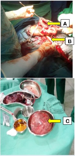
A midline longitudinal incision was made on the anterior abdominal wall to gain access to the peritoneal cavity. Intra-operatively, she had a clean peritoneal cavity, there was a huge anterior uterine wall fibroid mass that extended to the lower uterine segment, the fibroid mass later weighed 1.3 kg. A lower segment incision was made on the uterus, cutting just below the fibroid mass, and deepened to deliver the baby. She was delivered of a live female baby with Apgar score of 9 in both the first and fifth minutes and birth weight of 2.8 kg. Placenta and membranes were delivered completely. It was difficult to close the uterine incision because of the large fibroid mass at the upper margin. Foley catheter tourniquet was applied to the lower segment just below the incision and the fibroid mass was enucleated by both blunt and sharp dissection through the caesarean section incision (Figure 1). The dead spaces were obliterated in layers with vicryl 2. The caesarean section incision was closed in 2 layers with vicryl 2, the serosa was closed with vicryl 2/0 and the tourniquet removed. Adequate haemostasis was secured, however, an intraperitoneal drain was put in place. The anterior abdominal wall was closed in layers. The surgery lasted 1 hour 50 minutes. Estimated blood loss was 600 mls. She did not require blood transfusion. Her post-operative haemoglobin concentration was 9.8 g/dl. She made good recovery and was discharged on the 6th post-operation day.
Second case
A 39-year old Nigerian primigravida booked for antenatal care in our facility at 19 weeks + 4 days. She presented with an earlier ultrasound scan with findings of a singleton intrauterine fetus with coexisting multiple fibroids the largest measuring 9.5 cm X 7.0 cm. On examination, her symphysiofundal height was 27 cm and there were multiple irregular masses palpated on the uterus. She continued her regular antenatal clinic visit till gestational age of 36 weeks when a repeat obstetric ultrasound scan reported a viable intrauterine singleton in oblique lie with the head to the right iliac fossa and coexisting multiple fibroid masses with the largest measuring 10.8 x 7.5 cm and occupying the lower portion of the anterior uterine wall below the fetal head. She frequently complained of lower abdominal pain during the ante natal period. The placenta was posterofundal in location. She was counselled for elective caesarean delivery. She gave consent and she was booked for surgery at 38 weeks.
On the day of surgery, her pre-operative haemoglobin concentration was 11.5 g/dl. Her vital signs were a pulse rate of 80 beats per minute, full volume, regular and the blood pressure was 120/70 mmHg and her respiratory rate was 22 cycles per minute. Intravenous tranexemic acid 1 g was administered 10 minutes before the start of surgery. A midline longitudinal incision was made on the lower anterior abdominal wall to access the peritoneal cavity. A clean peritoneal cavity was noted. A transverse lower segment uterine incision was made on the uterus, cutting through the large fibroid mass, and deepened to deliver a live male neonate with Apgar score of 8 in the first minute and 9 in the fifth minute and birth weight of 2.9 kg. The placenta and membranes were completely delivered. The cut fibroid mass at the edges of the incisions were enucleated to allow for ease of closure of the caesarean incision. The dead spaces were closed with vicryl 2 sutures. The other fibroids were not removed as they were small and far from the caesarean section incision. The uterine incision was then repaired with vicryl 2 and haemostasis was properly secured. An intra-peritoneal drain was inserted but remained inactive. The anterior abdominal wall was closed in layers. The surgery lasted 1 hour 30 minutes. Estimated blood loss was 800 mls. She did not receive intraoperative blood transfusion. Her post-operative condition was satisfactory. Her post-operative haemoglobin concentration was 9.5 g/dl. She had an uneventful post-operative recovery and was discharged on the 5th day after surgery.
Discussion
Inevitable caesarean myomectomy is a caesarean myomectomy that is done so as to enable the completion of the caesarean section procedure. Here, the removal of the fibroids is unavoidable if the procedure of caesarean section must be completed. Our patients had inevitable caesarean myomectomy, it was practically impossible to repair the uterine incision without removing the fibroids at the edge of the incision in the lower uterine segment. Inability to gain access to the lower uterine segment is a justifiable reason for caesarean myomectomy [1]. Some cases of inevitable caesarean myomectomy can be because it was impossible to close the abdominal cavity without removing the fibroids [8]. Inevitable caesarean myomectomy can be done when there is partial avulsion of subserous fibroids. Traditionally, caesarean myomectomy has been discouraged because of the potential maternal morbidity and mortality that can occur when haemorrhage ensues. However, it is documented that in carefully selected patients, it can be done [6,7].
There is no evidence to suggest that future fertility and or subsequent pregnancy outcome is affected by caesarean myomectomy [13]. The advantage of a caesarean myomectomy is that it spares the patient the cost of future myomectomy and hospital admission [6,14,15]. Moreover the immediate post-partum uterus is better adapted to handle haemorrhage than at any other time [7]. Again, it has been opined that during pregnancy, enucleation of fibroids is easier as the tissues become softer [14]. Besides, in some situation, as seen in our patients, it is inevitable. Because the fibroids were along the edge of the uterine incision making it difficult to close the incision, we performed inevitable myomectomy during cesarean section without need for blood transfusion.
Zhao et al [10] conducted a retrospective study on women who had fibroids in pregnancy to evaluate the safety and feasibility of caesarean myomectomy. They compared those who had caesarean myomectomy and those who had caesarean section and interval myomectomy. They noted that postpartum haemorrhage, neonatal weight, fetal distress, and neonatal asphyxia showed no statistical significance. They concluded that caesarean myomectomy could be safe and feasible. The above study and some other studies [6,16-18] are of the view that caesarean myomectomy can be safely done by experienced surgeon in well selected patients. In these studies and in many cases of caesarean myomectomy, the proportion of subserous myomas was significantly higher than in myomectomies performed in non-pregnant women. In patients with pedunculated subserous myomas, achieving haemostasis is not commonly a challenge since the fibroid mass has a stalk which can be ligated to secure haemostasis.
If caesarean myomectomy should be contemplated, certain precautions should be taken to reduce the risk of haemorrhage and other complications. The procedure should be done by an experienced surgeon who can combine speed and accuracy with respect to delivery of the baby and control of bleeding. Again, there may be need for hysterectomy should there be intractable haemorrhage [1]. The choice of abdominal incision should be such as to give adequate exposure. As were done for our cases, the sub umbilical midline skin incision will be ideal because it provides opportunity for extending the incision should the need arise. Classical uterine incision may be necessary when access to the lower uterine segment is difficult [1]. Medications aimed at reducing the risk of bleeding should be given. Our patients received intravenous tranexamic acid just before the procedure and intravenous oxytocin at the delivery of the baby. Oxytocin should be administrated via intravenous or local injection. Furthermore, haemostatic tourniquet can be applied after delivery of the baby but before removal of the fibroids. This is to reduce the intraoperative blood loss [10]. In addition, the surgeon and anaesthetist should be prepared for prolonged surgery time, prolonged exposure to anesthesia and the possibility of significant blood loss. The procedure should be done in facilities where blood transfusion services are available since there may be need for massive blood transfusion.
Patient who had caesarean myomectomy should be monitored more closely in the post-partum period for any evidence of haemorrhage. Haemorrhage could be in the form of vaginal bleeding or intraperitoneal haemorrhage, hence vital signs monitoring as well as vaginal and abdominal examinations are necessary. To further monitor for any intraperitoneal bleeding, peritoneal drain can also be inserted as were done for our patients. Clear cut instructions should be given on the plan for the subsequent pregnancy with respect to the timing and mode of delivery taking into consideration the increased risk of uterine rupture. This is especially true if multiple incision were made including breaching the endometrial cavity. Such patients should be counselled on the need for possible elective caesarean section in future pregnancies [1].
Our patients were elderly primigravidae presenting with fibroids in pregnancy. They both had ante natal complications namely repeated episodes of abdominal pain and breech presentation in the first case. They both had intramural fibroids in the lower segment which necessitated caesarean section and also precluded closure of the uterine incisions without removal of the fibroids. Blood loss was reduced intra-operatively with tranexemic acid and oxytocin infusion which was continued for 12 hours post-operatively. The first case benefited from application of tourniquet however it was difficult to access below the fibroid mass in the second case. None of the patients required blood transfusion. They both had uneventful post-operative period and were discharged at 6th and 5th day post operation respectively.
Conclusion
Uterine fibroids are common among blacks and due to delayed childbearing they may be encountered frequently by obstetricians. The management is historically conservative due to fear of haemorrhage and caesarean hysterectomy however there are cases where caesarean myomectomy is inevitable and can be successfully done with good outcome. These cases show that myomectomy during caesarean section can be safely performed with good outcome in carefully selected cases.
Declaration of patient consent: The authors certify that they have obtained all appropriate patient consent. The patients have given the consent for their images and other clinical information to be reported in the journal. The patients understood that their names and initials will not be published and due efforts will be made to conceal their identity, but anonymity cannot be guaranteed.
References
- Eli S, Kalio D, Abam D, Onumbu K, Pepple D, et al. Emergency inevitable caesarean myomectomy, challenge to obstetrician/ gynaecologist: a case report. Niger J Med. 2017; 26: 185-187.
- Ezeama C, Ikechebelu J, Obiechina N, Ezeama N. Clinical Presentation of Uterine Fibroids in Nnewi, Nigeria: A 5-year Review. Ann Med Health Sci Res. 2012; 2: 114-118.
- Ikpeze OC, Nwosu OB. Features of uterine fibroids treated by abdominal myomectomy at Nnewi, Nigeria. J Obstet Gynaecol (Lahore). 1998; 18: 569-571.
- Isah AD, Adewole N, Agida ET, Omonua KI. A Five-Year Survey of Uterine Fibroids at a Nigerian Tertiary Hospital. Open J Obstet Gynecol. 2018; 8: 468-476.
- Eyong E, Okon O. Large Uterine Fibroids in Pregnancy with Successful Caesarean Myomectomy. Case Reports Obstet Gynaecol. 2020; 8880296.
- Senturk M, Polat M, Doğan O, Pulatoğlu Ç, Yardımcı O, Karakuş R, et al. Outcome of Cesarean Myomectomy: Is it a Safe Procedure? Geburtshilfe Frauenheilkd. 2017; 77: 1200-1207.
- Garg P, Bansal R. Cesarean myomectomy: a case report and review of the literature. J Med Case Rep. 2021; 1-4.
- Aksoy AN, Saracoglu KT, Aksoy M, Saracoglu A. Unavoidable myomectomy during cesarean section: a case report. Health (Irvine Calif). 2011; 3: 156-158.
- Fouzia Z, Imose I. Navigating through the maze of caesarean myomectomy: generating evidence. Int J Reprod Contracept Gynecol. 2019; 8: 4646-4653.
- Zhao R, Wang X, Zou L, Zhang W. Outcomes of Myomectomy at the Time of Cesarean Section among Pregnant Women with Uterine Fibroids : A Retrospective Cohort Study. Biomed Res Int. 2019; 2019: 1-7.
- Umezurike C, Feyi-waboso P. Successful myomectomy during pregnancy: A case report. Reprod Health. 2005; 2: 1-3.
- Ravi R, Ashimini V, Lopamudra B. Myomectomy in Pregnancy: feasibility and safety. Int J Reprod Contracept Gynecol. 2017; 6: 4204-4208.
- Adesiyun A, Ojabo A, Durosinlorun M. Fertility and obstetric outcome after caesarean myomectomy. J Obstet Gynaecol (Lahore). 2008; 28: 710-712.
- Kathpalia SK, Arora D, Vasudeva S, Singh S. Myomectomy at cesarean section: A safe option. Med J Armed Forces India. 2016; 72: S161–3.
- Chauhan A. Caesarean myomectomy: Necessity or Opportunity. J Obs Gynecol India. 2018; 68: 432-436.
- Parveen S, Noor N, Madan I, Kulsoom U. A case series of caesarean myomectomy. Int J Reprod Contraception, Obstet Gynaecol. 2021; 10: 3580-3583.
- Yeasmin S, Hasanat M, Saha E, Rahman M, Sunyal D. Safety and feasibility of myomectomy during caesarean myomectomy during caesarean section in case of pregnancy associated with fibroid uterus. Mediscope. 2018; 5: 5-9.
- Baflbu A, Esma Y, Yavuzcan A, Kaya AE, Göynümer FG. Myomectomy during cesarean section: is it a safe procedure ? Perinat J. 2018; 26: 112-116.
