Clinical & Medical Surgery
Open Access
Volume 2
Ka Man Karen Ng1; Man Wai Chow2; Lucci Lugee Liyeung2; Samuel Ka-Kin Ling3*; Patrick Shu-Hang Yung3
*Corresponding Author: Ling Ka Kin Samuel
Department of Orthopaedics and Traumatology, The Chinese University of Hong Kong, Hong Kong.
Tel: 35052010; Email: samuel.ling@link.cuhk.edu.hk
Article Info
Received: Nov 03, 2022
Accepted: Dec 02, 2022
Published: Dec 16, 2022
Archived: www.jclinmedsurgery.com
Copyright: © Samuel LLK (2022).
Abstract...
Regeneten is a bioinductive implant designed to promote tendon regeneration. Evidence shows promising results in rotator cuff tears with early data demonstrating potential promotion of tendon healing. However, studies on the application of this bioinductive implant elsewhere in the body is very limited. This is the first case report of using this bioinductive scaffold to augment an Achilles tendon repair.
Citation: KMK Ng, MW Chow, LL Liyeung, SKK Ling, PSH Yung. The Application of a Bioinductive Implant in Achilles Ten- don Repair: A Case Report. J Clin Med Surgery. 2022; 2(2): 1061.
Introduction
Achilles tendon is the largest and strongest tendon in the body, yet it is also the most commonly injured tendon. Veronesi et al has reported that Achilles tendon rupture accounts for 40% of all tendon ruptures with a male predominance [1]. Anatomical study done by Chen et al. showed that the midportion of the Achilles tendon was supplied by the peroneal artery and is markedly more hypovascular than the rest of the tendon, which was supplied by the posterior tibial artery [2]. As a result, together with a small cross-sectional area, most rupture occurs at around 2-6 cm proximal to the calcaneal insertion at midportion of the tendon.
Achilles tendon is composed of 20% sparse cells and 80% extracellular matrix, which consists of a network of type I collagen fibres arranged in parallel and regular fashion and they provides tensile strength to the tendon [3]. Achilles tendon demonstrated non-linear elasticity, which means that after being stretched beyond its physiological limit, the tendon will eventually break [4,5].
The common injury mechanisms causing Achilles tendon rupture includes unexpected ankle dorsiflexion, violent dorsiflexion of a plantar flexed ankle and pushing off the weight-bearing foot with an extended knee. There are severe risk factors contributing to Achilles tendon rupture, for example, obesity, history of local steroid injection and underlying tendinopathy.
Regarding the management of Achilles tendon rupture, many factors should be taken into consideration, including age and functional demand of patients, chronicity of injury, site of rupture and tendon status, i.e. any underlying tendinopathy. For older patients with low functional demand, conservative management with either cast immobilization or functional brace can be offered and a recent randomized controlled trial reported that there was no evidence to show that traditional plaster casting is superior to early weight-bearing in a functional brace [6]. For young and active patients, surgical repair is recommended either by open or minimally invasive approach. In case of pre-existing tendinosis or chronic tear, flexor hallucis longus tendon transfer, VY advancement flap or turn down flap might have to be performed as well.
Use of bioinductive implant, e.g. Regeneten, has shown promising result in treatment of rotator cuff tear but its application elsewhere is very limited. In this case report, we are going to report a case of Achilles tendon rupture in a young and active gentleman with underlying tendinosis using Regeneten for repair augmentation.
Case report
Here we report a 42-year-old gentleman who was an amateur football player and part-time coach presented to us with right Achilles tendon rupture. He had a history of left Achilles tendon rupture while playing football in 2017 and was treated with open repair at that time. He was however complicated by tendon re-rupture 3 months after surgery due to another injury and was again treated with open repair.
This time he presented to us with sudden onset sharp pain over right heel upon initiation of sprint during a football game with his right ankle in forced dorsiflexion. He was unable to bear weight afterwards. During his initial physical examination, there was no open wound nor any obvious bruising. There was a palpable 2 cm gap over the mid-portion of the right Achilles tendon with tenderness over both the proximal and distal stump ends. Thompson’s test was positive. Lateral view of the right ankle x-ray showed no associated fracture nor calcification along the Achilles tendon. Ultrasound scan of the right Achilles tendon confirmed the diagnosis of a complete Achilles tendon rupture 7 cm proximal to its calcaneal insertion and the torn fibres are retracted with a 0.9 cm gap with mild background tendinosis.
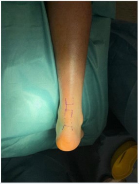
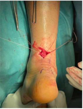
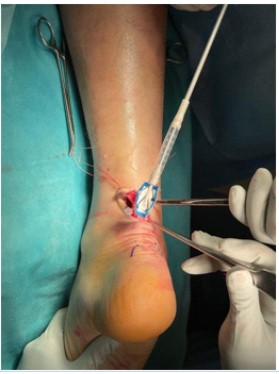
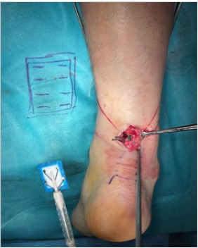
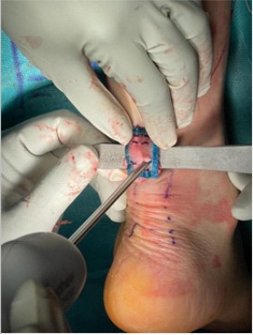
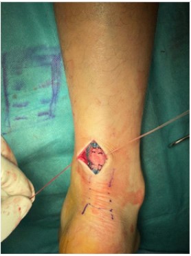
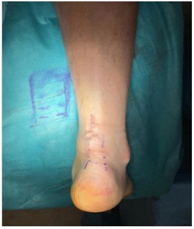
Patient was first given a dorsal block slab with right ankle in full plantarflexion after admission and open repair was arranged for him the next day. Operation was performed under general anaesthesia with the patient lying prone. Both stump ends of the ruptured right Achilles tendon were marked and then a 3 cm longitudinal skin incision was made along the medial border of the Achilles tendon. Paratenon was split in the middle longitudinally and both sides were tagged for repair later. Haematoma was then evacuated. Both stump ends were retrieved and were found to be flimsy. As a result, we first performed end-to-end direct repair with 2-strand core Orthocord suture in Krackow manner then augmented the site of repair with Regeneten. Regeneten was first being hydrated with sterile saline for approximately 2 minutes and after it had been fully deployed, it was placed on top of the repaired tendon and secured with 8 absorbable soft tissue staples. Paratenon was closed with 3-O Vicryl and skin was closed with 3-O Stratafix Monocryl. Our patient was discharged uneventfully 3 days after operation and the rehabilitation plan was as follows: non-weight bear walking in walker boot with two 10-degree heel wedges for 2 weeks then weight-bear walking as tolerated in the same boot for 2 weeks then weight-bear walking as tolerated in walking boot with one 10-degree heel wedge for 2 weeks then off walking boot and to start full weight bear walking.
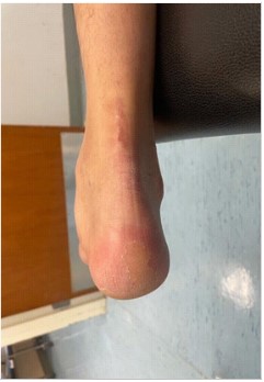
Our patient returned to the clinic for follow up on day 13 after operation. At that time he was able to non-weight bear walk with two elbow crutches and his wound condition was satisfactory. The repaired right Achilles tendon was intact and non-tender on palpation. Thompson’s test was negative and active right ankle plantar flexion has a muscle power grade of 4/5. At 6 week after the operation, our patient was able to full weight bear walk with two elbow crutches and reported no pain over the repaired tendon. The scar was well and the tendon was intact without any sign of infection. Active ankle dorsiflexion was up to plantigrade and plantarflexion was around 30 degrees. Power of both dorsiflexion and plantarflexion were grade 5/5. Patient was satisfied with his current condition.
Discussion & conclusion
Successful Achilles tendon repair can be a challenge to orthopaedic surgeons especially in mid-substance tear with poor local vascularity and in case with background tendinosis. According to previous studies, there are risk factors contributing to tendon re-rupture including male gender, underlying tendinopathy and high demand patients, etc. As our patient possessed all of these and with the history of tear and re-rupture of the contralateral side Achilles tendon, his condition warranted a more promising procedure then simply direct end-to-end repair alone.
Regeneten (Smith & Nephew) is a bioinductive implant derived from bovine Achilles tendon composed of type I collage and acts as a highly porous scaffold. It does not have a high tensile strength to provide structural support to the repaired tendon but it works to induce collagen formation and remodelling of the damaged tissue. It would be completely absorbed within 6 months after application.
Besides its use in repair of rotator cuff tear, there were only two case reports featuring the use of Regenten in treatment of partial patellar tendon and proximal hamstrings tendon tear [7,8]. In both cases, postoperative Magnetic Resonance Imaging (MRI) showed that there was improvement in tissue integrity and tendon thickness with absence of previous tendinosis and patients enjoyed a faster rehabilitation progress.
To our understanding, this is the first published case using Regeneten for Achilles tendon repair augmentation. There was no reported intra and postoperative complication so far. Our patient also enjoyed an enhanced rehabilitation without the need for casting after operation.
To further evaluate the long-term result, a plain MRI of the right Achilles tendon was arranged for him at 6 months after operation for review of the tendon healing status. Parameters including tendon thickness, tendon integrity and surrounding soft tissues status, etc. were of our research interest. Also other outcomes including the Visual Analogue Scale (VAS) for pain and the Foot and Ankle Outcome Score (FAOS) would also be evaluated in follow ups.
References
- Veronesi F, Borsari V, Contartese D, Xian J, Baldini N, et al. The clinical strategies for tendon repair with biomaterials: A review on rotator cuff and Achilles tendons. Journal of Biomedical Materials Research Part B: Applied Biomaterials. 2019; 108: 1826- 1843.
- Chen TM, Rozen WM, Pan W-ren, Ashton MW, Richardson MD, et al. The arterial anatomy of the Achilles tendon: Anatomical Study and Clinical Implications. Clinical Anatomy. 2009; 22: 377- 385.
- Zhu S, He Z, Ji L, Zhang W, Tong Y, et al. Advanced nanofiber-based scaffolds for Achilles tendon regenerative engineering. Frontiers in Bioengineering and Biotechnology. 2022; 10.
- Maganaris CN, Narici MV, Maffulli N. Biomechanics of the Achilles tendon. Disability and Rehabilitation. 2008; 30: 1542–1547.
- Morin C, Hellmich C, Nejim Z, Avril S. Fiber rearrangement and matrix compression in soft tissues: Multiscale hypoelasticity and application to tendon. Frontiers in Bioengineering and Biotechnology. 2021; 9
- Costa ML, Achten J, Marian IR, Dutton SJ, Lamb SE, et al. Plaster cast versus functional brace for non-surgical treatment of Achilles tendon rupture (UKSTAR): A multicentre randomised controlled trial and economic evaluation. The Lancet. 2020; 395: 441-448
- Looney AM, Fortier LM, Leider JD, Bryant BJ. Bioinductive collagen implant augmentation for the repair of chronic lower extremity tendinopathies: A report of two cases. Cureus. 2021; 13: e15567
- Mc Millan S, Faoao DO, Ford E. Treatment of an Insertional High Grade Partial Patellar Tendon Tear Utilizing a Bio-Inductive Implant. Arch Bone Jt Surg. 2019; 7: 203-208.
