Clinical & Medical Surgery
Open Access
Volume 2
*Corresponding Author: Swatishree Nayak
Department of Ophthalmology, AIIMS, Raipur, Chhattisgarh, India
Email: nswatishree@yahoo.com
Article Info
Received: Oct 25, 2022
Accepted: Nov 17, 2022
Published: Nov 24, 2022
Archived: www.jclinmedsurgery.com
Copyright: © Nayak S (2022).
Abstract...
Ocular surface squamous neoplasia is a broad entity that includes dysplastic lesions involving the squamous epithelium of conjunctiva and cornea. It is commonly seen in males between 50-75 years of age and has higher incidence in places close to the equator. Although a disease seen in 75% of cases unilaterally in older males, the younger cohort can have a bilateral presentation, where there is a strong suspicion of immunosuppression. Herein, we report a rare case of bilateral ocular surface squamous neoplasia in an immunocompetent patient.
Citation: Nayak S. A Rare Case of Bilateral Ocular Surface Squamous Neoplasia in an Immunocompetent Patient. J Clin Med Surgery. 2022; 2(2): 1060.
Introduction
Ocular Surface Squamous Neoplasia (OSSN) illustrates the abnormalities of epithelium of both conjunctiva and cornea as a whole. It embodies premalignant lesions like dysplasia, preinvasive carcinoma in situ and also invasive squamous cell carcinoma, a malignant lesion [1]. Generally, it is a slow growing tumor that seldom metastasizes but is capable of producing widespread local tissue destruction. The frequency of cases of OSSN documented worldwide is 0.02-3.5 cases per 100,000 people with countries located close to the equator having the maximum incidence [2,3]. However, currently there has been a changing trend with cases of OSSN rising in varied geographical locations like India. Incidence of bilateral OSSN worldwide is as low as 0.3% to 4% in those with no known systemic or local risk factors to as high as 11% to 15% in immunocompromised patients and 44% in patients with Xeroderma Pigmentosa [4]. In a recent study published from India, the prevalence and incidence of bilateral OSSN was 3% and 17 per 1000 cases of OSSN in 5 years respectively [5]. Bilateral OSSN is more commonly associated with immunosuppression and Xeroderma Pigmentosa. Herein, we report a rare case of bilateral OSSN in an immunocompetent patient.
Case report
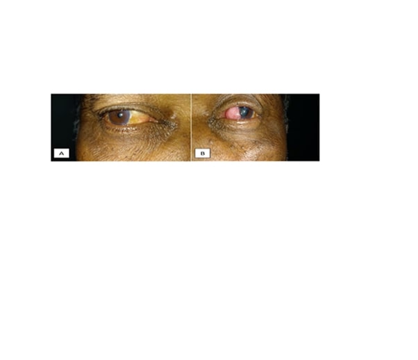
A healthy 65-year-old male presented to the Ophthalmology OPD with complaints of a painless, gradually increasing mass in his left eye for the last two and half years. The patient initially developed redness and foreign body sensation followed by development of an elevated lesion on the medial side of bulbar conjunctiva extending to the cornea which progressively increased in size. There was no previous history of trauma or surgery in that eye. He was a farmer by occupation and had a history of long exposure to sunlight during day time. He was diabetic, on oral hypoglycaemic agents for the past nine years and was a chronic smoker too. A detailed ophthalmic evaluation revealed best corrected visual acuity of 20/20 in right eye and 20/40 in left eye. On examination of left eye, a single, firm, immobile, painless strawberry-like shiny oval mass of 9 X 8 mm in size with well-defined margins was present over left nasal bulbar conjunctiva extending from caruncle up to limbus involving 7-10 o’clock position and encroaching 2-3 mm of cornea (Figure 1B). The lesion was elevated with an irregular surface and vascular core. Surprisingly, the right eye examination also revealed a 4 X 4 mm diffuse lesion over nasal bulbar conjunctiva with yellowish discolouration which mimicked pinguecula and had not been noticed by the patient (Figure 1A). Blood investigations were normal and serology test for Human Immunodeficiency virus (HIV) was negative. The clinical features in left eye were suggestive of OSSN.
Wide surgical excision remains the predominant modality of management of OSSN and further adjunctive treatment is based on histopathology reports. One week after presentation, the patient underwent left eye excisional biopsy of the mass by Shield’s no touch technique and cryo application to the dissected surface. The mass was easily dissected off the corneal surface and surface was covered with preserved Amniotic Membrane Graft (AMG) with cyanoacrylate glue.
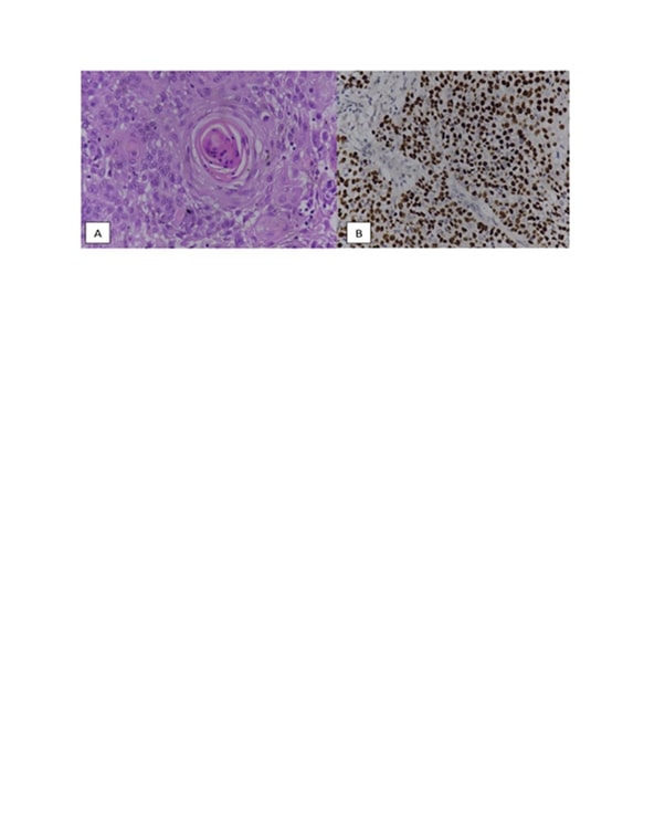
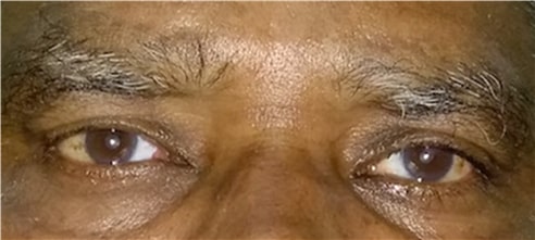
Histopathological analysis of specimen demonstrated breach in the basement membrane of basal epithelium and sheets of malignant cells showing pleomorphism, irregular nuclei, abundant eosinophilic cytoplasm with prominent nucleoli and keratin pearls leading to a diagnosis of moderately differentiated squamous cell carcinoma (Figure 2A). Immunohistochemistry assay revealed strong and diffuse p40 positivity of the tumour cells confirming their squamous origin (Figure 2B). Although the patient had no bothersome symptoms and right eye findings were completely incidental, right eye excisional biopsy of the lesion was done keeping in mind the probability of bilateral OSSN. Histopathology of the excised mass from right eye revealed focal moderate dysplasia and surface goblet cell changes, and hence, was considered as OSSN as well. Patient was kept on regular follow-up and there was no recurrence of the lesion even after two years (Figure 3).
Discussion
The term OSSN initially suggested by Lee and Herst in 1995 was classified into three categories encompassing all carcinomatous and dysplastic lesions of the ocular surface: 1) Benign dysplasia including papilloma, pseudotheliomatous hyperplasia, benign hereditary intraepithelial dyskeratosis 2) Preinvasive OSSN including conjunctival/ corneal carcinoma in situ and 3) Invasive OSSN including squamous carcinoma and mucoepidermoid carcinoma [6]. Geographical location has a definitive role in incidence of OSSN. This is clearly evident from the fact that annual incidence of OSSN in Australia, which is closer to the equator, is 1.9/100,000 population as compared to 0.03/100,000 population in the United States [7,8]. The incidence of invasive squamous cell carcinoma reported worldwide is even lesser, varying from 0.02 to 3.5/100,000 population [9]. Various risk factors predisposing to OSSN include ultraviolet light (UV) exposure, viral infections like Human Papilloma Virus and HIV, cigarette smoking, chronic inflammatory diseases of the ocular surface such as mucous membrane pemphigoid, chronic blepharoconjunctivitis, atopic eczema and vitamin A deficiency, defective DNA repair as in Xeroderma Pigmentosum [10]. Although a disease seen in 75% of cases unilaterally in older males, the younger cohort can have a bilateral presentation, where there is a strong suspicion of immunosuppression [11].
However, our patient was a 65-year-old male who was HIV seronegative. He was a farmer by occupation and hence exposure to UV rays together with smoking habits could have been the causative agent. UV rays, mostly UV-B rays cause significant direct DNA damage by crosslinking adjacent bases to create reactive oxygen species like hydroxyl radical, superoxide radical and cyclobutane pyrimidine dimers [12]. This disturbs genome stability and stem cell function and is the hallmark of carcinogenesis. OSSN classically presents as a unilateral vascularized limbal mass at the interpalpebral fissure [12]. Though clinical diagnosis is feasible, histopathology remains the gold standard for definitive diagnosis of OSSN, grading and also aids in deciding the treatment strategy [13]. In our case, histopathology findings of moderately differentiated squamous cell carcinoma in left eye led to a strong suspicion of OSSN in right eye too and prompted us to do an excisional biopsy of the diffuse lesion present in right eye.
The management of OSSN includes surgical resection, topical chemotherapy (mitomycin C, 5-fluorouracil), topical immunomodulation with interferon alpha-2b and photodynamic therapy [11]. As the lesion in our case involved only 3 clock hours of limbus, excisional biopsy using Shield’s no touch technique i.e. excision with 4 mm clinically clear margins was done. This was followed by application of cryo (double freeze thaw technique) to destroy the residual tumor cells if any. The resulting defect was then covered by cryopreserved amniotic membrane with cyanoacrylate glue. As the histopathological evaluation revealed tumor free margins, the patient was not administered any adjunctive therapy. Surgical excision of OSSN has a recurrence of 3.2%-67%, while the above multimodal approach reduces the recurrence rate to as low as 5% [14,15]. In our case, there has been no signs of recurrence at the two-year follow-up visit in both eyes.
Conclusion
Bilateral OSSN is rarely seen and when present, is commonly associated with immunosuppression. The novelty of this case was the presence of bilateral OSSN in an immunocompetent individual. While one eye had benign dysplasia, the other eye had invasive squamous cell carcinoma representing the two ends of the spectrum of OSSN. In conclusion, we would like to suggest that once histopathological diagnosis of OSSN is made, any suspicious lesion in the asymptomatic eye mandates evaluation and prompt management even in an immunocompetent individual. The malignant tendency of OSSN also makes it mandatory for the clinicians to regularly follow-up these patients.
References
- Basti A, Macsai MS. Ocular surface squamous neoplasia: A review. Cornea. 2003; 22: 687-704.
- Meel R, Dhiman R, Vanathi M, Pushker N, Tandon R, et al. Clinicodemographic profile and treatment outcome in patients of ocular surface squamous neoplasia. Indian J Ophthalmol. 2017; 65: 936-941.
- Gichuhi S, Sagoo MS, Weiss HA, Burton MJ. Epidemiology of ocular surface squamous neoplasia in Africa. Trop Med Int Health. 2013; 18: 1424-1443.
- Kaliki S, Kamal S, Fatima S. Ocular surface squamous neoplasia as the initial presenting sign of human immunodeficiency virus infection in 60 Asian Indian patients. Int Ophthalmol. 2017; 37: 1221-1228.
- Vempuluru V, Pattnaik M, Ghose N, Kaliki S. Bilateral ocular surface squamous neoplasia: A study of 25 patients and review of literature. Eur J Ophthalmol. 2022; 32: 620-627.
- Lee GA, Hirst LW. Ocular surface squamous neoplasia. Surv Ophthalmol. 1995; 39: 429-450.
- Lee GA, Hirst LW. Incidence of ocular surface epithelial dysplasia in metropolitan Brisbane. A 10-year survey. Arch Ophthalmol. 1992; 110: 525-527.
- Sun EC, Fears TR, Goedert JJ. Epidemiology of squamous cell conjunctival cancer. Cancer Epidemiol Biomarkers Prev. 1997; 6: 73-77.
- Tunc M, Char DH, Crawford B, Miller T. Intraepithelial and invasive squamous cell carcinoma of the conjunctiva: analysis of 60 cases. Br J Ophthalmol. 1999; 83: 98-103.
- Gichuhi S, Ohnuma S, Sagoo MS, Burton MJ. Pathophysiology of ocular surface squamous neoplasia. Exp Eye Res. 2014; 129: 172-182.
- Cicinelli MV, Marchese A, Bandello F, Modorati G. Clinical Management of Ocular Surface Squamous Neoplasia: A Review of the Current Evidence. Ophthalmol Ther. 2018; 7: 247-262.
- Das S, Nagesh N, Hegde R. Ocular surface squamous neoplasia. Current Indian Eye Research. 2019; 6: 4-16.
- Alomar TS, Nubile M, Lowe J, Dua HS. Corneal intraepithelial neoplasia: In vivo confocal microscopic study with histopathologic correlation. Am J Ophthalmol. 2011; 151: 238-247.
- Mirzayev I, Gündüz AK, Özalp Ateş FS, Özcan G, Işık MU. Factors affecting recurrence after surgical treatment in cases with ocular surface squamous neoplasia. Int J Ophthalmol. 2019; 12: 1426-1431.
- Honavar SG, Manjandavida FP. Tumors of the ocular surface: A review. Indian J Ophthalmol. 2015; 63: 187-203.


