Clinical & Medical Surgery
Open Access
Volume 2
*Corresponding Author: Nayan Bhindi
Department of Plastic & Faciomaxillary Surgery, The Alfred Hospital 55 Commercial Rd Melbourne VIC 3004, Australia.
Tel: 03-9076 0270; Email: nbhindi5@gmail.org.au
Article Info
Received: Sep 14, 2022
Accepted: Oct 19, 2022
Published: Oct 28, 2022
Archived: www.jclinmedsurgery.com
Copyright: © Bhindi N (2022).
Abstract...
Introduction: Debilitating pain from amputation stump neuromas is a difficult surgical problem. Targeted Muscle Reinnervation (TMR) is a recent surgical technique involving residual peripheral nerve stumps being coapted to a proximal target muscle motor point to improve myoelectric bioprostheses control. Here, we evaluate the efficacy of TMR of gastrocnemius at amputation to manage a symptomatic tibial nerve injury.
Methods: A 38-year-old, previously high-functioning, female presented for management of intractable nerve pain and deteriorating function following left tibial nerve injury. A below knee amputation (BKA) with TMR involving tibial nerve coaptation to motor fascicles of the gastrocnemius was performed. Primary endpoints included Visual Analogue Pain Scale (VAPS) and Short Form-36 (SF-36) scores measured preoperatively and 13 months postoperatively. Postoperative analgesia requirements formed a secondary outcome measure.
Results: Both pain and self-reported function displayed significant improvement 13 months postoperatively, resulting in significant reduction in post-operative analgesic requirement.
Conclusion: TMR may useful in treating chronic nerve-related pain once conservative methods fail. By restoring ‘physiological continuity’ of damaged peripheral nerves, primary TMR can reduce the incidence of symptomatic neuromas; improving both quality of life and prosthesis utilisation.
Citation: Bhindi N, Angliss M, Shayan R, Bruscino-Raiola F. Targeted Muscle Reinnervation of the Gastrocnemius for Preventing Neuropathic Pain. J Clin Med Surgery. 2022; 2(2): 1051.
Introduction
Neuroma formation is a seemingly inevitable consequence following nerve transection when regenerating axons are unable to re-enter the distal nerve segment [1]. Postamputation neuromas are a significant source of morbidity, causing intractable pain to limitations in functional capacity. It was estimated that 1.6 million people in the United States were living with limb amputations in the year 2005, with the prevalence predicted to more than double by 2050 [2]. Painful neuromas may develop in 13% to 32% of all major limb amputees even in the setting of a well-fitted prosthesis [3]. In cases of traumatic amputation, the incidence of residual limb neuroma pain has been reported as high as 71% [4].
A myriad of conservative and surgical treatments has been proposed for neuroma management, however, a single consistently effective strategy is currently lacking [1,3,5]. Targeted Muscle Reinnervation (TMR) is a decade-old surgical technique which has recently shown promise in managing neuromas recalcitrant to conventional methods. Originally indicated for providing myoelectric control of prostheses, TMR involves coaptation of the proximal nerve stump to an adjacent secondary motor point with a vascularised scaffold [4-6]. Here, we present our clinical experience with successfully treating a symptomatic tibial nerve injury using TMR of the gastrocnemius muscle.
Patient and methods
Informed consent was obtained from the patient discussed in this case study. A previously high-functioning 38-year-old-Caucasian female was referred to our centre for further surgical management of intractable nerve pain and deteriorating function following a kayaking-related trauma to her left leg four years prior. Initial injury involved the superficial posterior compartment of the leg, with a partial tear through the left gastrocnemius and plantar is muscles. The patient was conservatively managed using crutches for six months, and eight weeks post weight bearing the muscles tore further. Multiple repair surgeries were attempted, although complicated by the development of acute compartment syndrome, an unrecognised intra operative rupture of the tibial artery and later, chronic tibial nerve pain. Neurolysis of the tibial nerve extending from knee to ankle failed to relieve the nerve pain and ten days postoperatively a Staphylococcus aureus wound infection developed. The wound remained open and chronically non-healing for eight months requiring several hospital admissions for oral and intravenous antibiotics.
Almost two years since the initial injury, neurolysis and a right parascapular free flap was performed to temporise the on-going nerve pain. Despite initial improvements, the patient represented after two years with recurrence of nerve symptoms and declining ambulation. A decision was made to perform a semi-elective left trans-tibial or Below Knee Amputation (BKA) and TMR of the gastrocnemius muscle. A BKA was accepted by the patient to provide better function while the authors considered TMR offered the best chance of managing the refractory nerve pain. The patient was discharged on day 7 postoperatively following an uncomplicated stay and rehabilitation.
Operative technique
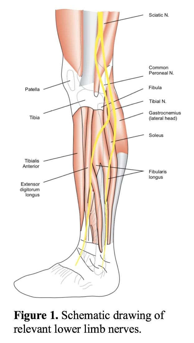
The procedure was conducted under general anaesthesia with short acting muscle relaxants. First stage comprised a left trans-tibial amputation approximated 10cm below the tibial tuberosity, due to the quality of the soft tissue. Upon identifying the tibial nerve (Figure 1), a neurotomy was performed proximal to the injured and scarred segment.
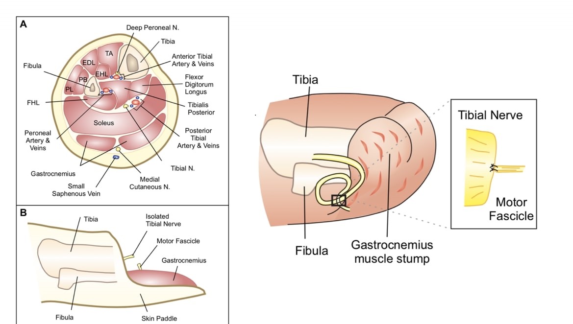
In the second stage, innervation to the gastrocnemius muscle was identified using nerve stimulation. A small nerve fascicle was dissected as close as possible to the muscle and subsequently divided. The proximal segment of this fascicle was hyper-innervated with the distal end of the tibial nerve using 8/0 nylon. Size discrepancy between the tibial nerve and fascicle was estimated 12:1. To prevent avulsion of the anastomosis, the epineurium of the tibial nerve was secured to the epimysium of the gastrocnemius muscle using 8/0 nylon (Figure 2).
Finally, closure of the soft tissue defect was achieved with a posteriorly based myocutaneous flap. The stump was closed in layers and a VAC dressing applied to splint and control the volume of the stump.
(Left) (A). Cross-sectional biew through amputation stump. (B). Lateral trans-tibial amputation stump view highlighting the posteriorly basked skin paddle and dissected nerves prior to coaptation. (Right) Insect of anastomosis between the tibial nerve and motor fascicle. Note: 12:1 size discrepancy.
Abbreviations: TA: Tibialis Anterior; EDL: Extensor Digitorum Longus; EHL: Extensor Hallucis Longus; PL: Peroneus Longus; PB: Peroneus Brevis.
Outcome assessment
Primary endpoints1. Visual Analogue Pain Scale (VAPS): a 10-point grading of VAPS was used to evaluate the patient’s severity of pain preoperative and postoperatively.
2. Short Form-36 (SF-36): was used to assess subjective self-reported level of function both preoperative and postoperatively. Scoring was performed using the RAND method.
Secondary endpoint
1. Preoperative and postoperative analgesia requirements were recorded.
Results
Comparative results between preoperative and postoperative primary endpoints of VAPS and SF-36 are shown in Table 1. Significant improvement in pain scores was demonstrated postoperatively using both VAPS and the pain component of SF-36. Over the last 3 months, the patient reported very brief episodes (~30 seconds) of nerve pain with a maximal VASP score of 8.
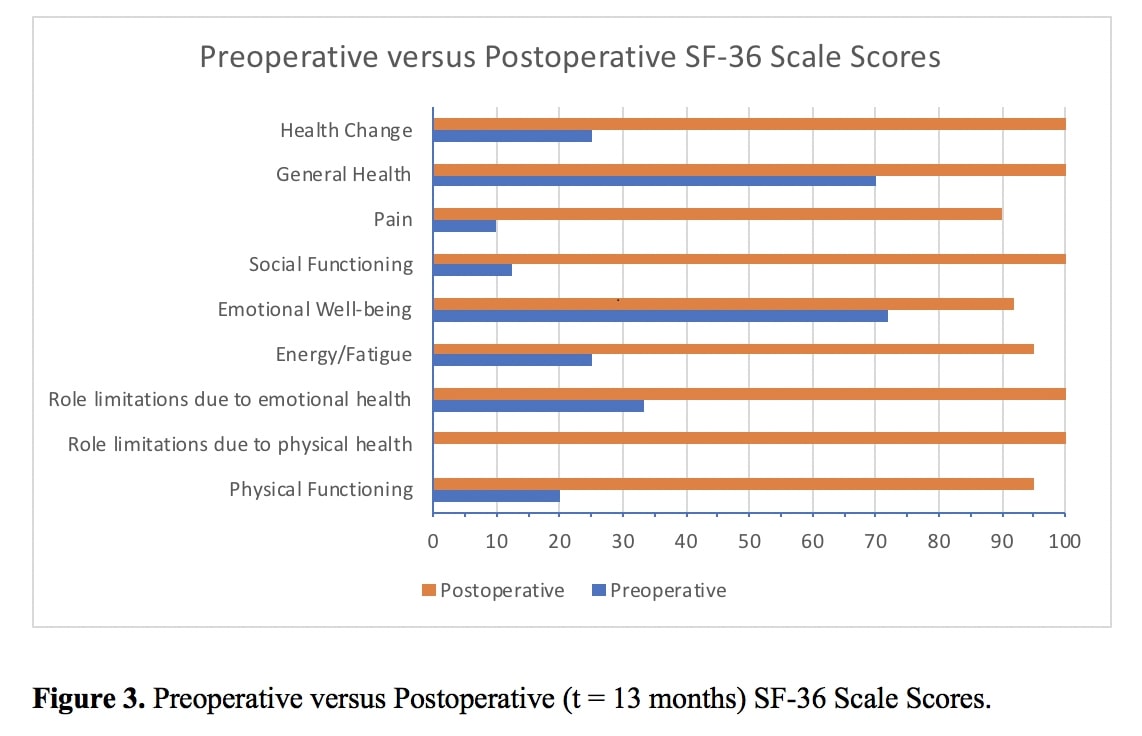
A global improvement of all eight domains of function in the SF-36 assessment was also achieved compared to the preoperative period (Figure 3). Pre-TMR the patient was unable to walk ≥50 m more than once/week and was reliant on crutches for ambulation. Since the intervention, she has been able to weight bear on her stump (via prosthesis) and perform to her self-reported premorbid level of function.
Discussion
Neuromasarise when the proximal segment of a transected peripheral nerve lacks a distal nerve target and/or in the absence of neurotrophic factors to guide regenerating axons [4]. A painful neuroma may be more troublesome to the patient than loss of motor function [1]. Often the emotional suffering of these patients is further compounded by multiple treatment failures aimed at relieving their chronic pain.
Table 1: Comparison of primary endpoint outcomes preoperatively and postoperatively (t = 13 months).
| Primary Endpoints | Preoperative Period | Postoperative Period |
|---|---|---|
| VAPS | 9 | 1 |
| SF-36 scores | ||
| Physical Functioning | 20 | 95 |
| Role Limitations - Physical Health | 0 | 100 |
| Role Limitations - Emotional Health | 33.33 | 100 |
| Energy/Fatigue | 25 | 95 |
| Emotional Wellbeing | 72 | 92 |
| Yes (n=123) | n=97 (56%) | n=26 (17%) |
| Social Functioning | 12.5 | 100 |
| Yes (n=116) | n=95 (54%) | n=21 (14%) |
| Pain | 10 | 90 |
| General Health | 70 | 100 |
VAPS: Visual Analogue Pain Scale; SF-36: short form-36.
Table 2: Comparison of preoperative and postoperative (t = 13 months) analgesic requirements.
| Preoperative Period | Postoperative Period |
|---|---|
| Pregabalin 75 mg BD | Amitriptyline 20 mg nocte |
| Tramadol SR 100 mg BD | |
| Tramadol IR 50 mg PRN | |
| Targin 5-10 mg nocte | |
| Endone 5 mg PRN |
SR: Sustained Release; IR: Immediate Release.
The optimal surgical technique for preventing neuromas is widely debated. Sharp resection, either with cautery or scalpel, while placing gentle traction on the nerve and allowing retraction of the transected end into the proximal region of the limb is one of the most commonly used techniques in recent years [7]. It is believed that retraction of the transected segment into surrounding muscle tissue creates a protected micro environment, rendering neuroma formation less favourable.
Despite recognition of the problem posed by chronic stump pain, current treatments are unsatisfactory with variable efficacy. Conservative strategies are broadly classified into pharmacological, psychological, and physical methods. First line therapy typically comprises narcotics and nonsteroidal anti-inflammatory drugs, with the potential for adjuvant agents such as antidepressants, anticonvulsants, lidocaine patches, and even, steroid injections and nerve blocks. Psychological treatment entails mirror box therapy while physical methods may include transcutaneous or spinal electrical nerve stimulation, rehabilitation with exercise or massage [3]. Although non-curative, conservative avenues should be explored thoroughly prior to subjecting the patient to potentially unwarranted surgical or anaesthetic risks.
Greater than 150 surgical methods have been proposed for the treatment of symptomatic neuromas [6,8]. Techniques have ranged from simple external neurolysis, traction neurectomy, silicone nerve capping, centro central anastomoses, to more complex nerve transpositions into proximal tissues and coverage with pedicled or free flaps [9,10]. The lack of a single definite surgical therapy stems from the inconsistency of current methods. However, a study comparing surgical therapies in intact upper limb neuromas suggested superior outcomes were achieved with techniques attempting to reconstitute the peripheral nervous system anatomy over other methods [8].
Here, the authors justified novel use of TMR for the treatment of chronic nerve pain given the failure of other modalities. TMR restores the physiological continuity of a damaged nerve and although other techniques, such as nerve grafting or use of vein conduits, are guided by similar principles, their postoperative clinical course have been unpredictable. Findings from this case validate the resolution of chronic amputation nerve pain demonstrated retrospectively in a significant number of patients where TMR was indicated for intuitive control of bioprostheses [4-6]. Marked improvement in both primary and secondary endpoints during the postoperative period has lead the authors to hypothesise that TMR may prevent neuroma formation by enabling organised axonal regeneration. Post-surgical imaging and histologic studies are required to prove this assertion and exclude the possibility of asymptomatic neuroma recurrence. Noting the limitations of this single-centre experience, the authors believe primary TMR (at the time of amputation) should be considered as a serious candidate for managing recalcitrant nerve pain, although extensive clinical trials with long-term follow-up are required for a definitive conclusion.
Conclusion
Our experience confirms that patients with chronic nerve pain refractory to non operative methods and reduced functional capacity can significantly benefit from TMR. By allowing sprouting axons somewhere to go and something to do, physiological continuity of the transected segment is restored, thus preventing the formation of a disorganised symptomatic neuroma.
Conflict of interests/financial disclosures:
NoneReferences
- Vernadakis AJ, Koch H, Mackinnon SE. Management of neuromas. Clin Plast Surg. 2003; 30: 247-268
- Ziegler-Graham K, MacKenzie EJ, Ephraim PL, Travison TG, Brookmeyer R. Estimating the prevalence of limb loss in the United States: 2005 to 2050. Arch Phys Med Rehabil. 2008; 89: 422-429.
- Ducic I, Mesbahi AN, Attinger CE, Graw K. The role of peripheral nerve surgery in the treatment of chronic pain associated with amputation stumps. Plast Reconstr Surg. 2008; 121: 908-914.
- Souza JM, Cheesborough JE, Ko JH, Cho MS, Kuiken TA, et al. Targeted muscle reinnervation: a novel approach to postamputation neuroma pain. ClinOrthopRelat Res. 2014; 472: 2984-2990.
- Pet MA, Ko JH, Friedly JL, Mourad PD, Smith DG. Does targeted nerve implantation reduce neuroma pain in amputees? Clin Orthop. 2014; 472: 2991-3001.
- Bowen JB, Wee CE, Kalik J, Valerio IL. Targeted Muscle Reinnervation to Improve Pain, Prosthetic Tolerance, and Bioprosthetic Outcomes in the Amputee. Adv Wound Care (New Rochelle). 2017; 6: 261-267.
- Whipple RR, Unsell RS. Treatment of painful neuromas. The Orthopedic clinics of North America. 1988; 19: 175-185
- Guse DM, Moran SL. Outcomes of the surgical treatment of peripheral neuromas of the hand and forearm: a 25-year comparative outcome study. Ann Plast Surg. 2013; 71: 654-658.
- Mackinnon SE, Dellon AL, Hudson AR, Hunter DA. Alteration of neuroma formation by manipulation of its microenvironment. Plast Reconstr Surg. 1985; 76: 345-353.
- Wu J, Chiu DT. Painful neuromas: a review of treatment modalities. Ann Plast Surg. 1999; 43: 661-667.


